The Mystery of the Pigmented Plaque on the Chin
Sabrina Oujdi*, Hanane Baybay, Siham Boularbah, Sara Elloudi, Meryem Soughi, Zakia Douhi and Fatima Zahra Mernissi
Department of Dermatology, University Hospital Hassan II, Morocco
Received Date: 19/08/2023; Published Date: 04/01/2024
*Corresponding author: Dr Sabrina Oujdi, Department of Dermatology, University Hospital Hassan II, Fes, Morocco
Abstract
The atrophoderma of pierini and pasini is a rare form of morphea which is characterized by plates of atrophic emble and which affects most often the trunk, we reported a case of pierini pasini localized at the level of the face in the form of pigmented plate in a young woman.
Keywords: Atrophoderma of Pasini and Pierini; Morphee; Chin
Introduction
Pigmentations of the face and neck are the most cosmetically important and constitute a very frequent reason for consultation; they are frequent in middle-aged women and melasma is the most frequent cause.
Observation
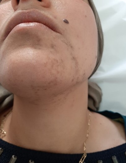
A 30-year-old female patient with no notable pathological history consulted for asymptomatic pigmented lesions on the face that had been evolving for 18 months (Figure 1).
On clinical examination: a poorly defined pigmented plaque was noted, roughly linear and slightly atrophic, on the left half of the chin, reaching the left perioral area, with no other cutaneous or mucosal lesions
The evoked diagnoses were: lichen pigmentogen and atrophoderma of pierini and pasini
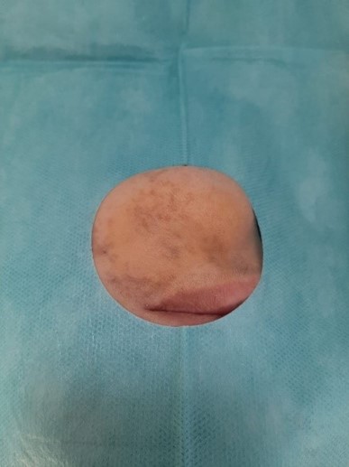
The skin biopsy (Figure 2) revealed a skin tissue covered by a regular and atrophic epidermis with hyperpigmentation of the basal layer; the underlying dermis was the site of a moderate mononuclear inflammatory infiltrate, essentially perivascular, and the skin appendages were preserved in favor of pierini and pasini atrophoderma.

Figure 1: Clinical image of pigmented plaque on the chin.

Figure 2: biopsy site on the cutaneous side of the lower lip
Discussion
Pierini and Pasini Atrophoderma (PPA) is a rare entity that usually affects young adults with a female predominance.
The cause of atrophoderma of Pasini and Pierini remains unknown. Some authors have linked it to infection with Borrelia burgdorferi [1] and some others have suspected a neurological origin due to the zoster-like appearance of the lesions, but no clear evidence has been found in relation to this hypothesis [2].
The usual clinical presentation is one or more atrophic plaques of brown or purplish color without inflammation or associated sclerosis, it is currently part of the forms of morphea but what differs from it is the absence of the lilac ring in the initial phase clinically [3].
PPA is mainly located on the trunk and lower limbs. The evolution is slow over several years. These lesions stabilize for 10 to 20 years. Spontaneous regression has been described. Histologically, skin biopsy of healthy and injured skin shows epidermal atrophy with hyperpigmentation of the basal layer, a perivascular lymphocytic infiltrate and homogenization of collagen bundles in the deep dermis. The cutaneous appendages and the elastic network remain preserved.
Among the differential diagnoses we note Moulin's linear atrophoderma which is a rare dermatosis defined by the presence of unilateral, atrophic and hyperpigmented lesions arranged according to Blaschko's lines rarely localized on the face as well; the histology is often non-specific, it can show a hyperpigmentation of the basal layer of the epidermis, a perivascular lymphocytic infiltrate of the dermis and an ascension of the sweat glands; the main distinctive criterion is that AAP never follow on the Blashko lines [4,5].
A Lebanese review published in 2008 that included 16 patients with pierini and pasini atrophoderma found that only 2 patients with associated lesions on the rest of the body had this pathology on the face and underlined the rarity of this pathology in this location [6].
The therapeutical approach also includes topical corticosteroids and antimalarials, Furthermore, topical treatments using calcineurin inhibitors were also reported, although with variable responses.
Conclusion
Pierini and Pasini's Atrophoderma (PPA) is a dermatosis initially described by Pasini in 1923 and Pierini in 1936. It is a rare dermatosis whose etiopathogeny remains controversial, we reported the case of an APP of atypical location
Consent
The examination of the patient was conducted according to the principles of the Declaration of Helsinki.
The authors certify that they have obtained all appropriate patient consent forms, in which the patients gave their consent for images and other clinical information to be included in the journal. The patients understand that their names and initials will not be published and due effort will be made to conceal their identity, but that anonymity cannot be guaranteed.
Conflict of Interest: None
References
- González-Morán A, Martín-López R, Ramos ML, Román C, González-Asensio MP. [Idiopathic atrophoderma of Pasini and Pierini. Study of 4 cases]. Proceedings Dermosifiliogr, 2005; 96(5): 303-306.
- Bassi A, et al. Idiopathic congenital atrophoderma of Pasini and Pierini. Arch Dis Child, 2015; 100(12): 1184.
- Litaiem N, Idoudi S. Atrophoderma of Pasini and Pierini. 2022 Aug 8. In: StatPearls [Internet]. Treasure Island (FL): StatPearls Publishing, 2023. PMID: 30085611.
- Lahouel I, et al, the atrophoderma of Pierini and Pasini: about three cases Author links open overlay panel,1016/j.revmed.2020.10.252.
- Saleh Z, et al, Atrophoderma of Pasini and Pierini: a clinical and histopathological study. Journal of Cutaneous Pathology, 2008; 35(12): 1108–1114.
- Charfi O, et al. Dermatology, Charles-Nicolle Hospital, Tunis, Tunisia 2 Department of Internal Medicine a, Charles-Nicolle Hospital, Tunis, Tunisia 3 Department of Internal Medicine b, CHU Charles-Nicolle, Tunis, Tunisia 4 Anatomical pathology, Charles-Nicolle Hospital, Tunis, Tunisia.
- Pope E, Laxer RM. Diagnosis and management of morphea and lichen sclerosus and atrophicus in children. Pediatr Clin North Am, 2014; 61(2): 309-319.

