Pachydermoperiostosis - A Case Report and its Surgical Management
Anantheswar YN Yelambalase Rao, Harish Kabilan*, Krithika Jagadish, Srikanth Vasudevan, Ashok Basur Chandrappa, Dinkar Sreekumar and Annika Marwah
Department of Plastic surgery Manipal Hospital, India
Received Date: 01/06/2023; Published Date: 04/10/2023
*Corresponding author: Harish Kabilan, Department of Plastic surgery Manipal Hospital, India
Abstract
Pachydermoperiostosis (PDP), is an autosomal-dominant/autosomal recessive inherited disorder. PDP goes by various other names such as Rosenfeld-Kloepfer or Touraine–Solente–Gole syndrome, and primary or Idiopathic Hypertrophic osteoarthropathy) PDP is characterized by the coarsening of facial features, with furrowing of the forehead and scalp regions, new periosteal bone formation, arthralgia, and digital clubbing. The medical management is well reported in literature along with the histopathological and genetic basis of the disease. However, the approach to cosmetic deformity in the form of forehead furrows is rarely reported. A case report of a young adult male is presented here. His chief complaint at the time of presentation was the deepened furrows over his forehead and the prematurely aged look on his face. Here we describe our surgical approach and the outcome of the same.
Introduction
PDP was first described in 1868 by Friedreichin, who called it ‘Hyperostosis of the entire skeleton [1].
Unna in 1907 then coined the term ‘cutis verticis gyrata’ for the thick, folded skin over the scalp and forehead [2].
It was in 1935 that a dermatologist named Touraine recognized this condition as a familial disorder and then classified it into three forms: complete (with pachyderma and periostosis), incomplete (without pachyderma) and the forme fruste (Minimal skeletal changes with pachydermia) [3]. idiopathic or Primary Hypertrophic osteoarthropathy is more commonly known as pachydermoperiostosis (PDP) and is classically defined by major and minor criteria. Pachydermia, periostosis, and digital clubbing are the major criteria and the minor criteria include cutis verticis gyrata, seborrhea with sebaceous hyperplasia, folliculitis, hyperhidrosis, and acne.
Approximately 95% of cases are secondary HOA and 5% are primary HOA [4].
Below we report the case of a patient with the complete form of primary HOA (ie, PDP), who came to us with complaints of deepened furrows on the forehead and an aged appearance. He had been previously investigated & treated for Hansen’s disease.
Case Description
A male patient, aged 26 years, unmarried, a pharmacist by profession. Of note in his medical history was that his parents were consanguineously married. He had undergone a laparoscopic appendectomy when he was 16 years old. He also has an elder brother who underwent a renal transplant (father donor) for a congenital renal issue (exact details unknown).
His treatment history included azathioprine, and prednisolone, by the rheumatology
Department for management of pain in his fingers, toes, and knees; and history of botox for management of his forehead furrows more than 6 months before presentation.
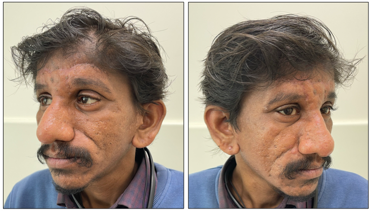
Local examination of his face revealed multiple deepened horizontal furrows running across the forehead, along with deepened vertical glabellar furrows. He also had deep bilateral nasolabial folds and thickened skin over his entire face.
Radiographic studies of the skull, long bones, spine, pelvis, hands, and feet revealed hyperostosis of the forearm bones, the femur, and the tibia, and hyperostosis of the metatarsal bones, osteophytes, and sclerosis in the right foot.
Several folds (cutis verticis gyrata) were also present in the occipital region, which weren’t noticeable due to his hair. Palmar skin was rough and his fingers and toes showed evidence of clubbing, and the toes in addition had thickened nails.

Figure 1 (A, B, C, D): Preoperative pictures of the patient with very pronounced folds in the area of the forehead, between the eyes, and in the nasolabial grooves. Thickened eyelid edges are also evident, and the patient’s general facial expression appears to be sad. Moreover, the chief complaint of this 26-year-old male was that he looked older than he was.

Figure 2
A. X-RAY HAND AP; OBL Alignment is normal. Carpal, metacarpal, phalanges and the joints are normal. No periarticular demineralization or erosions. No soft tissue thickening. No fracture or dislocation.
B. X-RAY WRIST BOTH AP, LATERAL Carpals are normal in alignment and are normal. Joint spaces are preserved. Known Pachydermoperiostosis, consolidated periosteal reaction in the lower ends of radius and ulnar and metacarpals causing undertubulation and widening, no bone destruction.
C. X-RAY BOTH FOOT AP, LAT Known Pachydermoperiostosis Diffuse symmetric widening of the metatarsals. No erosions. Tarsal; TM and MP and IP joints do not show erosions. No increased soft tissue density. No fracture or dislocation.
D. Evidence of clubbing in the right toes.
E. X-RAY BOTH ANKLE AP, LAT Ankle joint is normal in alignment. Subtaloid joint does not show any abnormality. Known Pachydermoperiostosis, the consolidated shaggy periosteal reaction in the lower ends of tibia and fibula causing undertubulation and widening, no bone destruction
Routine investigations, as well as radiological investigations such as chest radiographs and ECG results, revealed no other abnormalities.
Surgical Procedure
After informed consent, the patient was taken up for surgery. The surgical procedure was as follows.
For the folds or furrows on the forehead, rhytidectomy was performed by elliptical excision and then redundant skin was removed (Figure 3 A).
The fibrous adhesions causing the furrows were adherent from the dermis up to the frontalis muscle fascia.
Excessive thickened skin and tissue were excised and the remaining skin was then scored vertically along the inner surface up to the dermis (Figure 3 B).

Figure 3
A - Preoperative marking of the elliptical incision
B - Vertical scoring of the inner surface
C - Mini facelift
D - Immediate post-op picture
The skin over the long vertical glabellar furrows were thinned, stretched and anchored laterally to the frontalis muscle fascia to reduce their prominence.
The forehead incision was sutured primarily in layers over a glove drain.
The bilaterally prominent nasolabial creases were addressed through a mini facelift and SMAS was plicated with 3-0 prolene
Lipofill injections were also planned for the nasolabial creases but could not be done due to inadequate fat availability in the donor areas due to the asthenic disposition of the patient.
After concluding the procedure, the final closure was done in layers with monocryl and prolene.
The patient developed mild wound dehiscence over the forehead suture line as well as over the left facelift suture line; both of which were managed conservatively.

Figure 4: Post-op results.

Figure 5: Pre and post-operative photographs for comparison.

Figure 6: The histopathological findings.
Discussion
Pachydermoperiostosis is a rare inherited disorder described by Touraine, Solente, and Golé in 1935, who proposed a classification of PDP characterized by thickened skin, periostosis, hyperhidrosis, and digital clubbing of both toes and fingers [5,6]. They found that the onset was typically around adolescent years, but the skin and bone changes worsen for 5 to 20 years and then cease around middle age. The aesthetic changes remain for a lifetime though [7-9].
Steroids [10], isotretinoin [11], colchicine [12], and botulinum toxin [13] are a few of the various modalities that have been tried for treating the cutaneous manifestations.
The aesthetic surgeries for PDP that have been documented in the literature include bilateral blepharoplasties [14], tarsal wedge resections [15], frontal rhytidectomy [16,17], along with facelifts [18]. The most evident and troubling complaint of the patient is the forehead creases, which first of all manifest prematurely and leave the patient looking distressed as well as aged. The scalp furrows even though deeper and is usually camouflaged by hair.
In our case study, we performed a direct rhytidectomy as well as additional surgical relaxation of the skin to lessen the skin tension, especially in the upper forehead and scalp. In PDP, the epidermis is hyperkeratotic with a thickened dermis. Although the use of retinoic acid and FU in the early stages may help slow down the process of epidermal proliferation and thickening for a delayed presentation like in our patient it is better to surgically reduce the thickness of the skin by thinning the skin and to score the inner surface of the dermis to allow for better pliability and uniformity.
A mini facelift was also performed bilaterally to alleviate and reduce the depth of the nasolabial grooves. The excess skin was removed and the SMAS was anchored to the zygomatic fascia. It was due to this submuscular and subfascial dissection and removal of redundant skin that forehead furrows were corrected.
Lipofill was deferred due to inadequate donor sites.
Cases of tissue expansion and forehead lift using endotine fixation devices for forehead suspension and brow elevation have been reported in the literature. This is considered superior to the single suture technique [19], thus leaving room for improvement in the future.
Conclusion
The prevalence of Pachydermoperiostosis is estimated to be 0.16%, with a male: female ratio of 7:1 [20].
Since the condition is mistaken to be mere facial rhytids or osteoarthritis, the clinical diagnosis is imperative to establish the condition before any further treatment decision is made.
Even after conclusive diagnosis, the classical approach so far has been drug therapy, with incomplete resolution and unsatisfactory results at least when it comes to the aesthetic aspect.
The surgical approaches for the aesthetic predicament in PDP - as limited as they may be in literature - lean towards a multimodality/ combination approach consisting of direct rhytidectomy, patch padding, mask lift, or fixed devices such as endotine fixation devices. In conclusion, this rare condition requires a comprehensive surgical approach that should be tailored to the specific needs of the patient.
References
- Friedreich N. Hyperostose des gesammten Skelettes. Arch Für Pathol Anat Physiol Für Klin Med, 1868; 43(1): 83-87. doi:10.1007/BF02117271
- UNNA P. Cutis verticis gyrata. Monatsh Prakt Derm, 1907; 45: 227-233.
- TOURAINE A. Un syndrome osteodermopathique : la pachydermie plicaturee avec pachyperiostose des extremites. Presse Med, 1935; 43: 1820-1824.
- Pachydermoperiostosis. NORD (National Organization for Rare Disorders), 2021.
- Supradeeptha C, Shandilya SM, Vikram Reddy K, Satyaprasad J. Pachydermoperiostosis - a case report of complete form and literature review. J Clin Orthop Trauma, 2014; 5(1): 27-32. doi: 10.1016/j.jcot.2014.02.003
- Chander R, Kakkar S, Jain A, Barara M, Agarwal K, Varghese B. Complete form of pachydermoperiostosis: a case report. Dermatol Online J, 2013; 19(2): 10.
- Rajan TMS, Sreekumar NC, Sarita S, Thushara KR. Touraine Solente Gole syndrome: The elephant skin disease. Indian J Plast Surg Off Publ Assoc Plast Surg India, 2013; 46(3): 577-580. doi:10.4103/0970-0358.122025
- Yao Q, Altman RD, Brahn E. Periostitis and hypertrophic pulmonary osteoarthropathy: report of 2 cases and review of the literature. Semin Arthritis Rheum, 2009; 38(6): 458-466. doi: 10.1016/j.semarthrit.2008.07.001
- Pachydermoperiostosis: an update - Castori - 2005 - Clinical Genetics - Wiley Online Library, 2021.
- Tanaka H, Maehama S, Imanaka F, et al. Pachydermoperiostosis with myelofibrosis and anemia: report of a case of anemia of multifactorial causes and its improvement with steroid pulse and iron therapy. Jpn J Med. 1991; 30(1): 73-80. doi: 10.2169/internalmedicine1962.30.73
- Cutis verticis gyrata and pachydermoperiostosis. Several cases in a same family. Initial results of the treatment of pachyderma with isotretinoin, 2021.
- Matucci-Cerinic M, Fattorini L, Gerini G, et al. Colchicine treatment in a case of pachydermoperiostosis with acroosteolysis. Rheumatol Int, 1988; 8(4): 185-188. doi: 10.1007/BF00270458
- Ghosn S, Uthman I, Dahdah M, Kibbi AG, Rubeiz N. Treatment of pachydermoperiostosis pachydermia with botulinum toxin type A. J Am Acad Dermatol, 2010; 63(6): 1036-1041. doi: 10.1016/j.jaad.2009.08.067
- Berdia J, Tsai FF, Liang J, Shinder R. Pachydermoperiostosis: a rare cause of marked blepharoptosis and floppy eyelid syndrome. Orbit Amst Neth, 2013; 32(4): 266-269. doi:10.3109/01676830.2013.788672
- Kumar S, Sidhu S, Mahajan BB. Touraine-Soulente-Golé Syndrome: A Rare Case Report and Review of the Literature. Ann Dermatol, 2013; 25(3): 352-355. doi: 10.5021/ad.2013.25.3.352
- Alaya Z, Boussofara L, Bouzaouache M, Amri D, Zaghouani H, Bouajina E. Complete form pachydermoperiostosis in Tunisia – A case series and literature review. Egypt Rheumatol, 2018; 40(2): 127-130. doi: 10.1016/j.ejr.2017.06.006
- Zhang Z, Zhang C, Zhang Z. Primary hypertrophic osteoarthropathy: an update. Front Med, 2013; 7(1): 60-64. doi:10.1007/s11684-013-0246-6
- RBCP - Surgical treatment of primary pachydermoperiostosis: report of two cases, 2021.
- Madruga Dias JAC, Rosa RS, Perpétuo I, et al. Pachydermoperiostosis in an African patient caused by a Chinese/Japanese SLCO2A1 mutation-case report and review of literature. Semin Arthritis Rheum, 2014; 43(4): 566-569. doi: 10.1016/j.semarthrit.2013.07.015
- Primary hypertrophic osteoarthropathy (pachydermoperiostosis). Report of 2 familial cases and literature review - ScienceDirect, 2021.

