Delivery After Cloaca Malformation Surgery
Rosita Aniuliene1, Povilas Aniulis1,* and Gilvydas Verkauskas2
1Department of Surgery, Lithuanian University of Health Sciences, Italy
2Department of Surgery, Vilnius University, Italy
Received Date: 04/05/2023; Published Date: 22/08/2023
*Corresponding author: Povilas Aniulis, Department of Surgery, Lithuanian University of Health Sciences, Italy
Abstract
Cloacal malformation It is extremely rare, occuring in approximately 1 in 25 000 births. We describe here a 19 years old patient who concieved naturally with cloacal malformation after surgery of cloaca and proctoplastica (propter atresia ani et recti), after surgery of vaginal septum with agenesis of right tube and ovary, agenesis of coccygeus, dysplasia sacri, aneurysma septi interatrialis, hydronephrosis,chronical pyelonephritis, fecal incontinence. In childhood patient underwent many recostructive surgeries. During pregnancy phlebothrombosis of v.femoralis communis, v.femoris profundi and v.iliaca externa occurred. A birth plan was discussed and approved together with paediatric surgeons and urologists. At 38 weeks of pregnancy, the patient was urgently hospitalized and underwent Caesarean section with cooperation with urologists. Newborn was delivered by cephalic presentation with birth weight of 2720 g and with an Apgar score of 9 and 9.
Introduction
Cloacal malformation is an anatomic defect occuring exclusively in females. It arises during the fifth to eight weeks of fetal development as a failure of the urorectal septum to form and divide the cloaca into rectum dorsally and urogenital sinus ventrally. This results in confluence of the rectum, vagina,and urethra into a single common channel. It is extremely rare, occuring in approximately 1 in 25 000 births. The etiology of persistent cloaca is unknown [1].
Persistent urogenital sinus (PUGS) is a pathological condition characterized by an abnormal communication between the urethra and vagina. It may be a part of a complex syndrome and can be more often associated with congenital malformations affecting the genitourinary tract system (33%) such as intersex, rectovaginal communication, bladder agenesis, absence of vagina, and hydrocolpos [2].
Cloacal malformation frequently has associated Mullerian anomalies with high incidence of gynecological problems that require surgical correction, such as hematometra due to cervical stenosis or vaginal stenosis, vaginal septum,and endometriosis [3].
20% of those patients suffering urinary incontinence and 40% fecal incontinence, sometimes-sexual dysfunction. About 80% of patients with cloacal malformation are born with a structural abnormality of the urinary tract, including renal dysplasia, ectopic kidney, solitary or duplex kidney, or ureteropelvic junction obstruction [4] and sacrum and spine anomalies [5].
We describe here a 19 years old patient who concieved naturally with cloacal malformation after surgery of cloaca and proctoplastica (propter atresia ani et recti), after surgery of vaginal septum with agenesis of right tube and ovary, agenesis of coccygeus, dysplasia sacri, aneurysma septi interatrialis, hydronephrosis,chronical pyelonephritis, fecal incontinence. During pregnancy phlebothrombosis of v.femoralis communis, v.femoris profundi and v.iliaca externa occurred.
Case Report
A 19-year-old patient at 34 weeks of pregnancy presented to the Gynaecological outpatient clinic of the Hospital of Lithuanian University of Health Sciences (HLUHS) Kaunas Clinics with diagnosis of phlebothrombosis in the left leg. The patient could not remember the last menstrual period and did not visit the Gynaecological outpatient clinic. The findings of Ultrasound revealed viable foetus at 34 weeks and 6 days.
Anamnesis: the patient was born with congenital dysplasia – Cloaca malformation-persistent urogenital sinus, rectal and anal atresia, caudal agenesis, sacral agenesis and atrial septal aneurysm.
On the second day after birth a double-barrel colostomy was carried out, since proctoplasty and an integrity of the large intestine will be restored in future.
In addition, bilateral ureterohydronephosis, which was more prominent on the right side, was found, thus percutaneous nephrostomy was performed.
Due to rectal and anal atresia, proctoplasty was performed by the Pen’s method when the patient was 11 months old (18/08/1998).
The aim of Pen’s surgery method – to draw the end of the large intestine to the abdominal wall and to form the anus with normal anal sphincter function [6].
At the age of 2 years OLD (22/09/1999), the patient was urgently hospitalized due to progressive bilateral hydronephrosis to the Pediatric Surgery. Cystogram showed grades IV- V right unilateral vesicoureteral reflux (VUR), hydroureter, and left nephrosclerosis due to the stricture at the intramural portion of the ureter and the pressure of constantly full bladder.
The decision was made to perform vesicostomy as an alternative to epicystostomy, however, after consultation with urologists from the USA and failure to perform cystoscopy, vesicostomy was postponed.
Folowing 3 months, cystoscopy was successfully performed, and the diagnosis of urogenital sinus was made. The patient underwent vesicostomy. Since the girl showed persistent dysfunction of the right kidney and bilateral nephrosclerosis, in July, 2003 (at the age of 6 years), ureteroileocutaneostomy (Bricker conduit) in the right lower quadrant was performed and vesicostomy closed.
The aim of surgery – to form urinary reservoir using a portion of intestine. The urinary reservoir collects urine from both ureters and drains it via stoma created in the abdominal wall [7].
After her first menstruations, at age 12, the girl was hospitalised due to persistent urogenital sinus: two orifices of the vaginas opened at the cervix of the urinary bladder. The patient again underwent surgery, during which urogenital sinus was mobilized, double vaginas disconnected, and after forming one channel, the distal vaginal portion was formed using patches from vagina tissue and urogenital sinus, and the urethra was tubularized. The vagina was dilatated with Hegar dilator of size 16Ch and after 3 months to size 20Ch.
After 5 months following the last operation, the patient had augmentation cystoplasty using a segment of ileum; the earlier uretercutaneous segment was removed due to nutritional disorder, and a new ureterovesical junction formed. Also, continent appendicovesicostomy was formed, and the girl was taught to use catheter via stoma every 2-4 hours using 12 Fr Nelaton type catheter.
At the age of 15, following 3 years of vaginoplasty and the urethra formation, the patient was taught to use catheter via the urethra, however, after almost two years, catherization again was started through stoma due to insufficient drainage of the bladder and hydronephrosis progression.
The patient at 34 weeks of pregnancy presented to the Obstetrics department of the (HLUHS) Kaunas Clinics. Blood tests (full blood count (FBC), electrolytes, kidney function) were within normal limits, apart from slight anaemia, blood test showed the presence of white blood cells, unremarkable level of red blood cells and proteins.
The findings of ultrasound revealed viable foetus at 34 weeks and 6 days, amniotic fluid and foetoplacental circulation were normal.
A birth plan was discussed and approved together with paediatric surgeons and urologists. Due to phlebothrombosis, fraxiparine 0.6 ml twice per day was administered, urinary tract infection was treated with cefuroxime 500 mg daily, and anaemia - by tardyferon 80 mg orally twice per day. The patient was advised to continue treatment after leaving the hospital, and if labour does not start earlier - to arrive at hospital at 38 weeks of pregnancy for Caesarean section (CS).
At 38 weeks of pregnancy, with the persistence of diarrhea for two days, the patient was urgently hospitalized to the Obstetrics department of (HLUHS) Kaunas Clinics. Acute pyelonephritis was diagnosed and urine culture showed a significant level of E.coli. Coagulogram showed a mild hypercoagulation. HIV and syphilis were negative.
The patient complained of increasing backache and she did not experience uterine contractions (UC). Cardiotocography (CTG) showed UC are every 3-7 minutes. After 2 hours, when the patient started experiencing regular UC 3 times per 10 minutes, the decision was made to start CS earlier with participation of urologist using endotracheal anaesthesia.
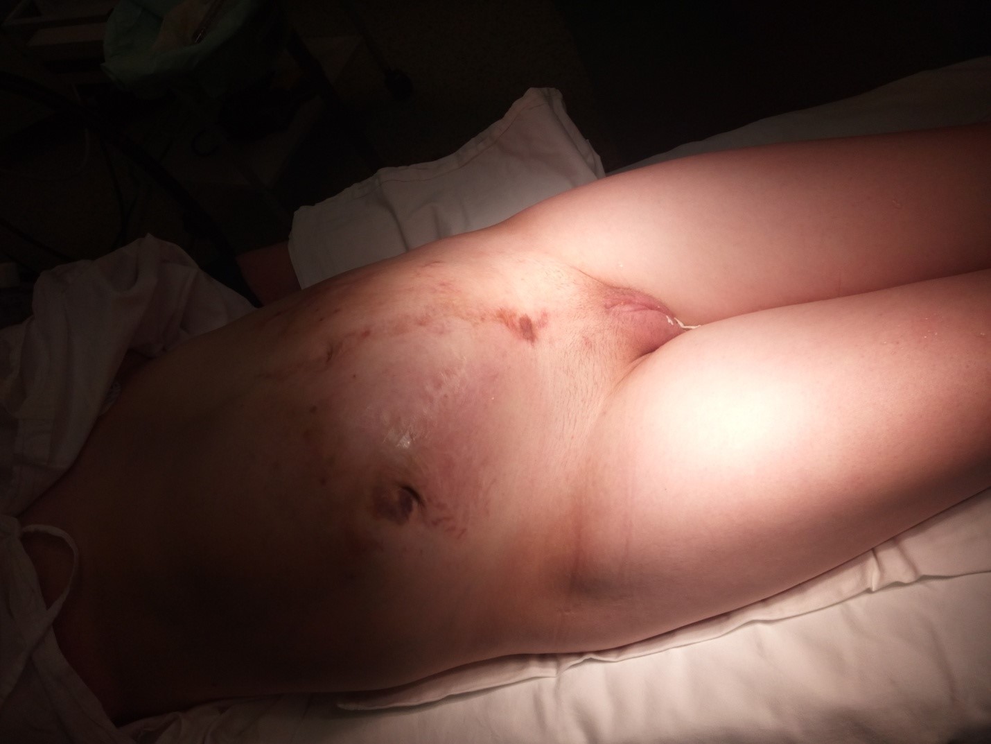
Before operation, the bladder was filled with fluid by Foley catheter and examined using ultrasound: the urine bladder was detected in the left lower quadrant (Figure 1, 2).
The abdominal cavity was incised by longitudinal section by passing the umbilicus from the left side. The incision length was 3- 4 cm superior the umbilicus and 7 cm superior the pubic bone.
The augmented bladder was filled by methylene blue using Nelaton type catheter.
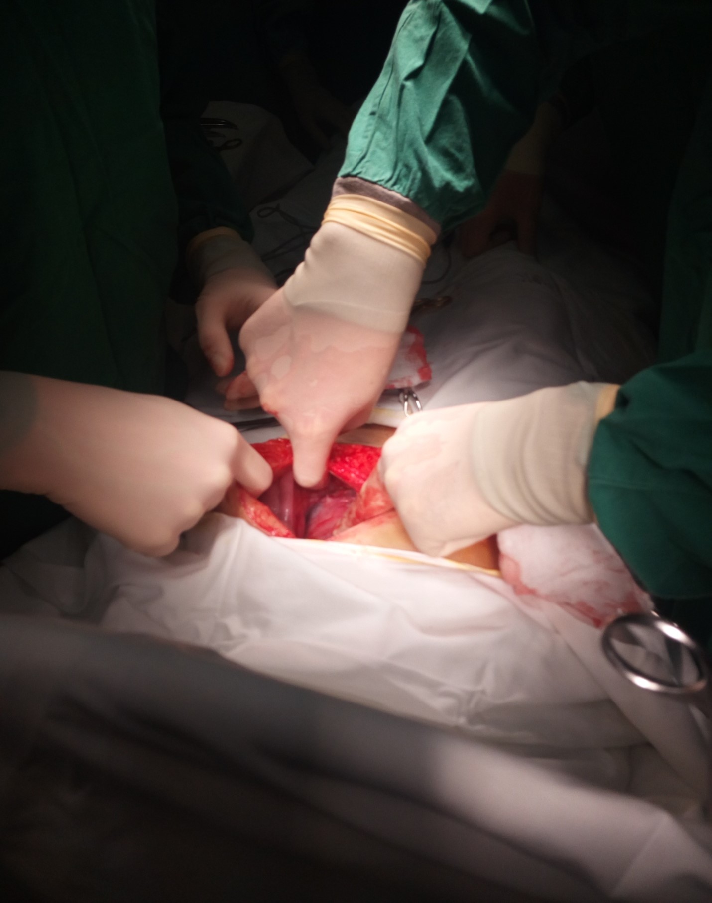
Adhesions between the uterus, peritoneum and omentum were divided using a speculum and dully pushing away the formed bladder to the left (Figure 3). A transverse uterine incision was made and female newborn was delivered by cephalic presentation with birth weight of 2720 g and with an Apgar score of 9 and 9.
The examination during surgery revealed absent of left adnexa.
Nelaton catheter inserted via stoma was left in the augmented bladder after surgery. Also, two drains remained in the pouch of Douglas and in the vesicouterine excavation.
Postoperative urine culture detected E.coli resistant to ampicillin, cefuroxime and nitrofurantoin. Cefuroxime was replaced by sulbactam 1.5 g 4 times per day.
During treatment, the primary healing of incision was observed, the patient was not febrile, a repeat urine test showed no changes, urine culture – sterile. The newborn was breast fed.
On the 10 day the patient was discharged with a healthy newborn from the hospital home.
Clinical Diagnosis: Grav. I- 38 hebd. Partus maturus. Dysplasia multiplex. Status post cloacae, atresiam ani et recti (proctoplasticam). Agenesia coccygis, dysplasia sacri. Agenesia ovarii et tubae dextri. Aneurysma septi interatrialis. Status post phlebotrombosam v. Femoralis communis, v. Profundae femoris, v. Iliacae externae. Enterocolitis acuta. Pyelonephritis chr. exacerbata. Sectio caesarea.

Figure 1: Ultrasound before operation.

Figure 2: Patient before s/c operation. On the right side-apendicovesicostoma.

Figure 3: The begining of operation-adhesiolysis.Žemiau ir on the left –gimda.Dešinėje- iš žarnos suformuotas šlapimo rezervuaras.
Discussion
Given the limited information on the pteferred mode of delivery of patients with PUGS repair.There have been several case reports of vaginal deliveries in these patients [8,9].
The patients who conceive after repairs invariably have had cesarean deliveries on the assumption that vaginal delivery would be difficult and/or dangerous. However, because they usually have undergone multiple previuos abdominal procedures, CS delivery also may carry greater risks than in the general population [10].
Alon Shrim,Tiina Podymow et al. from Canada described a patient with cloacal malformation, solitary kidney,bilateral fallopian tube obstruction, and didelphic uterus who reqiured in vitro fertilization to conceive and care ful surveilance resulted in an excellent pregnancy outcome with term delivery. Careful surveillance during pregnancy and a planned abdominal delivery resulted in a successful outcome [11].
The patients with cloacal malformations must give birth at third level university hospitals, because they need multidiscipline team for assistance.
References
- Warne SA, Wilcox DT, Creighton S, Ransley PG. Long-term gynecological outcome of patients with persistent cloaca. J Urol, 2003; 170(4Pt2): 1493-1496.
- Valentini AL, Giuliani M, Laino M, Zecchi V, Ninivaggi V, Manzoni C, et al. Persistent Urogenital Sinus: Diagnostic Imaging for Clinical Management.What Does Radiologist Need to Know? AmJ Perinatol, 2016; 33(5): 425-432.
- Warne SA, Wilcox DT, Ledermann SE, Ransley PG. Renal outcome in patients with cloaca. J Urol, 2002; 167: 2548-2551.
- Breech L. Gynecologic concerns in patients with anorectal malformations. Semin Pediatr Surg, 2010; 19: 139-145.
- Lund DP, Hendren WH. Cloacal exstrophy: a 25-year experience with 50 cases. J Pediatr Surg, 2001; 36(1): 68-75.
- Mathias A, Hutson JM. Anorectal Malformations in Children– Embryology, Diagnosis, Surgical management. Springer, 2006; 223-229.
- Smith DR, Galante M. The use of the bricker operation in urology. The American journal of surgery, 1987; 254-263.
- Greenberg JA, Hendren WH. Vaginal delivery after cloacal malformation repair.Obstet Gynecol, 1997; 90(4Pt2): 666-667.
- Ljubic A, Sulovic V, Stankovic A, Cvetkovic A. Cloacal dysgenesis and vaginal delivery ( article in French).J Gynecol Obstet Biol Reprod (Paris), 1993; 22: 417-418.
- Greenberg JA, Hendren WH. Vaginal delivery after cloacal malformation repair.Obstet Gynecol. 1997; 90(4Pt2): 666-667.
- Alon Shrim, Tiina Podymow, Lesley Breech, Michael H.Dahan. Term delivery after in vitro fertilization in a patient with cloacal malformation. J Obstet Gynaecol Can, 2011; 33(9): 952-954.

