Superficial Acral Fibromyxoma 2nd toe: A Rare Entity
Saumya Gupta1, Shreya Bhardwaj1, Pratibha2, Navneet Mishra3, Pranav Kumar Dave4,* and Vandana Agrawal5
1Resident Surgery, L N Medical College and J K Hospital, India
2Assistant Professor Surgery, L N Medical College and J K Hospital, India
3Department of Surgery, L N Medical College and J K Hospital, India
4Department of Radiology, L N Medical College and J K Hospital, India
5Department of Pathology, L N Medical College and J K Hospital, India
Received Date: 01/04/2023; Published Date: 10/07/2023
*Corresponding author: Dr Pranav K Dave, Prof. Deptt. of Radiology, L N Medical College and J K Hospital, E-7/540, MIG Senior, Arera Colony, Bhopal-462042, MP, India
Introduction
Superficial Acral Fibromyoma is a benign and rare tumour of the soft tissues. It’s also called digital fibromyoma and was first described by Fetsch in 2001, as a localized tumour in the acral extremities. Superficial acral fibromyoma often presents on either the fingers or toes, and has been shown to involve the nail in 97% of fingers, and 96% of toes [1]. Incidence of acral fibromyoma is not noted in the literature, and because of an unusual diagnosis, surgeons should be aware of this tumour, which requires complete surgical excision and follow up for recurrence. Since they are generally painless, intervention is late and a history of trauma is described in several cases.
Case Report
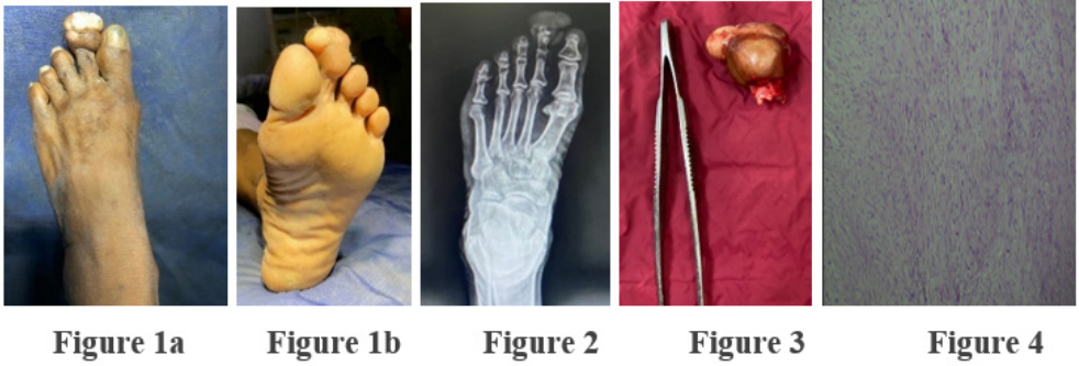
A 48-year-old healthy man presented with an asymptomatic, slowly growing nodule with a 15-year history on the tip of his second right toe. Examinations revealed a hemisphere, irregular-shaped growth, measuring 3.0 cm × 2.3 cm × 1.5 cm in size, on the tip of his second right toe (Figure 1). The nail was not visible and the nail bed was distorted. X ray right leg AP and lateral view was suggestive of excess of soft tissue components distally in the 2nd toe, bones and joints appeared normal (Figure 2). The USG local site was suggestive of lobulated echogenic mass lesion in the descending phalanx of the 2nd toe. Cytology was suggestive of scattered benign oval to spindle cells with no evidence of malignant cells. The growth was completely excised, with DIP disarticulation (Figure 3).

Histopathologically, the section showed dermal and subcutaneous tumour composed of bland spindle and stellate cells within myxoid and collagenous stroma. Scattered small calibre blood vessels and mast cells are seen. Cells are arranged in a random loose pattern and in focal areas forming short fascicles. No mitosis was seen. No nuclear atypia was noted (Figure 4).
Discussion
Superficial acral fibromyxoma is a benign and slow-growing solitary soft-tissue neoplasm [2]. The often-asymptomatic presentation of acral fibromyxomas means that there is often a delay in their diagnosis and subsequent treatment [3]. SAFM, a rare slow growing myxoid tumor in the subungual area that was first described in 2001 [4]. Clinically, neurofibroma, dermatofibrosarcoma protuberans (DFSP), low grade fibromyxoid sarcoma, and myxofibrosarcoma should all be considered in the differential diagnosis and evaluation of SAF [5]. It is also called digital fibromyxoma. It has a slight male predominance. The mean age of the patients falls in the 5th decade with patients ranging from 4 to 86 years of age. About 25% of cases recur [1,6].
It usually manifests itself through a painless mass of slow growth that affects mainly males in the fifth decade of life. It usually affects the distal region, with a polypoid or dome-shaped appearance [7]. The differential diagnosis of superficial acral fibromyxoma includes fibroma of the tendon sheath, myxoid neurofibroma, glomus tumor, giant cell tumor of the tendon sheath, sclerosing perineuroma, acral fibrokeratoma, cutaneous myxoma, myxoinflammatory acral fibroblastic sarcoma, fibrous histiocytoma, and dermatofibrosarcoma protuberans [8].
In conclusion, if a soft tissue tumor occurs under the nail, we should suspect superficial acral fibromyxoma and we also should keep in mind that such tumors can grow aggressively. Awareness of this entity is helpful in distinguishing SAF from other myxoid soft tissue tumors occurring there. Complete excision with clear resection margins is the mainstay of treatment [9].
References
- Hollmann TJ, Bovée JVMG, Fletcher CDM. Digital fibromyxoma (superficial acral fibromyxoma): a detailed characterization of 124 cases. Am J SurgPathol, 2012; 36(6): 789–798.
- Akçay Çelik M, Erdem H, TurhanHaktanır N. Superficial Acral Fibromyxoma: A case report. Int J Surg Case Rep, 2020; 77: 531-533. doi: 10.1016/j.ijscr.2020.11.048.
- Sivasaththivel M, Howard MD, Yazdabadi A. Acral fibromyxoma: a rare plantar nodule. BMJ Case Reports CP, 2022;15: e247565.
- Ramya C, Nayak C, Tambe S. Superficial Acral Fibromyxoma. Indian J Dermatol, 2016; 61(4): 457-459. doi: 10.4103/0019-5154.185734.
- DeFroda SF, Starr A, Katarincic JA. Superficial acral fibromyxoma: A case report. J Orthop, 2016; 14(1): 23-25. doi: 0.1016/j.jor.2016.10.018.
- Al-Daraji WI, Miettinen M. Superficial acral fibromyxoma: a clinicopathological analysis of 32 tumors including 4 in the heel, J. Cutan. Pathol, 2008; 35: 1020–1026.
- Crepaldi BE, Soares RD, Silveira FD, Taira RI, Hirakawa CK, Matsumoto MH. Superficial Acral Fibromyxoma: Literature Review. Rev Bras Ortop (Sao Paulo), 2019; 54(5): 491-496. doi: 10.1016/j.rbo.2017.10.011.
- Hashimoto K, Nishimura S, Oka N, Tanaka H, Kakinoki R, Akagi M. Aggressive superficial acral fibromyxoma of the great toe: A case report and mini-review of the literature. Mol Clin Oncol, 2018; 9(3): 310-314. doi: 10.3892/mco.2018.1669.
- Wang QF, Pu Y, Wu YY, Wang J. Superficial acral fibromyxoma of finger: report of a case with review of literature. Zhonghua Bing Li Xue Za Zhi, 2009; 38(10): 682-685.

