When HEMOPHILIA Comes Late: A Spontaneous Psoas Hematoma Revealing Acquired Hemophilia A
Ouaddane Alami R*, El Allali A, Ahsaini M, Mellas S, El Ammari J, Tazi MF, El Fassi MJ and Farih MH
Department of urology, Hassan II University Hospital, University Sidi Mohammed Ben Abdellah, Morocco
Received Date: 10/03/2023; Published Date: 25/05/2023
*Corresponding author: Rhyan Ouaddane Alami, Medicine doctor, Department of urology, Hassan II University Hospital, University Sidi Mohammed Ben Abdellah, Fez, Morocco
Abstract
Spontaneous psoas hematoma is a serious bleeding event in patients with bleeding disorders such as hemophilia. However, psoas hematoma is the source of costly treatment and can be the cause of complications. Among these, compression of the rural nerve is the most frequent and occurs in large-volume hematomas. Transformations into pseudotumor and myositis ossificans are fortunately rare in Western countries where treatment with clotting factors is available. Relapse is also frequently reported, often in the context of insufficiently prolonged treatment.
Keywords: Hemophilia; Psoas hematoma; Lombo-cruralgia
Introduction
Spontaneous psoas hematoma is rare, and often associated with a particular condition: anticoagulants, dialysis, systemic diseases. It represents a surgical emergency. The diagnosis must be suspected in case of cruralgia, or a sudden pain in the iliac fossa. There is generally no traumatic context; most hematomas arise spontaneously.
The clinical examination looks for a posits, more rarely a pain caused by deep palpation of the iliac fossa, which can radiate towards the loins. The reference examination to establish the diagnosis in an emergency abdominal-pelvic CT scan without injection (spontaneous hyper density within the psoas muscle). A complete blood count looks for deglobalization, that can be significant, and a complete hemostasis assessment can diagnose an unknown coagulopathy.
The management of spontaneous psoas hematoma depends on the severity of the bleeding, the hemodynamic status, and the neurological deficit. Conservative attitude with bed rest and correction of bleeding abnormalities, is usually sufficient in case of small hematoma, absence of active bleeding, or mild neurological symptoms. Surgical intervention is mandatory in case of severe motor dysfunction or hemorrhagic shock. Embolization offers a therapeutic alternative for hemodynamically unstable patients or patients undergoing high-risk surgery. With the increased use of anticoagulant, spontaneous psoas hematoma has become a more common condition and should not be overlooked. The major complication, which makes this pathology so serious, is damage of the femoral nerve compressed by the hematoma.
Case Report
A 50-year-old patient, with no specific history, who had presented a left lumbar pain (VAS: 9) for 10 days with sudden onset and progressive installation associated with motor deficit of the ipsilateral inferior member.
The clinical examination found an isolated motor deficit of the left quadriceps and hamstrings rated 1/5, associated with decreased sensitivity on the antero-internal face of the thigh with ecchymotic spots on left lumbar fossa.
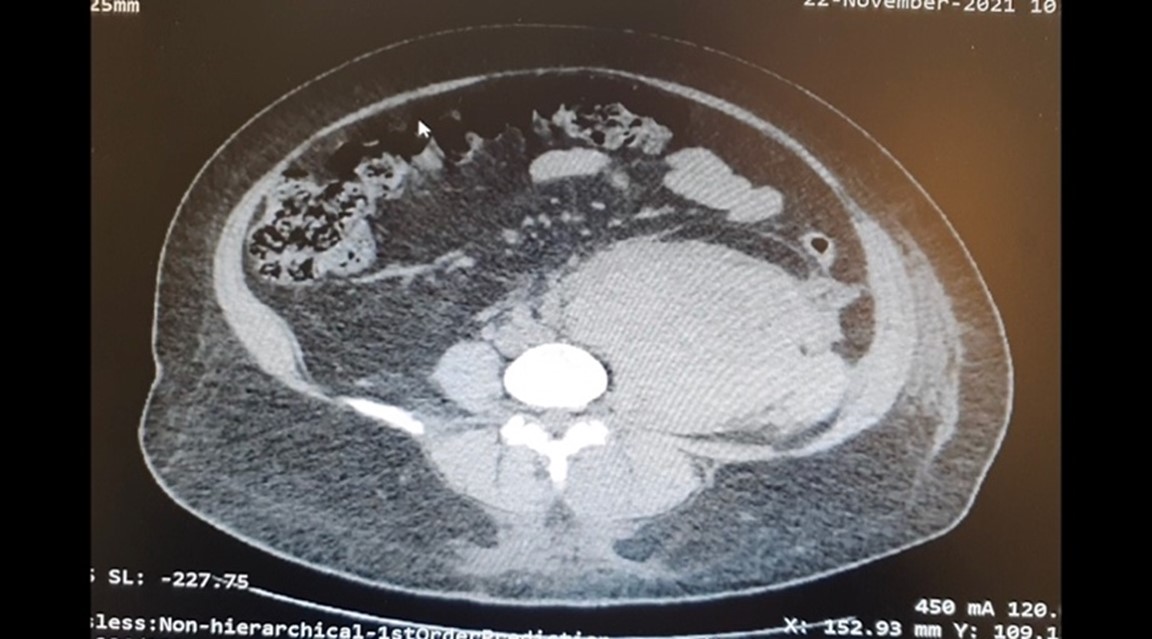
The patient initially admitted to a private clinic, where a complete biological assessment was carried out, which objectified an aPTT elongated to 69.6s, a 5,1g/dL of hemoglobin. an injected abdominal CT scan was performed, that has showed the presence of a large hematoma on left psoas muscle, measuring 18cm*12cm compressing the ipsilateral ureter with ureterohydronephrosis (Figures 1). The patient was then transferred to our structure for further care.
Initial management was to transfuse the patient with red blood cells and fresh frozen plasma, put some analgesic treatment with bi-antibiotic therapy based on third-generation cephalosporins and aminoglycosides to avoid infection of the hematoma.
We then completed our biological assessment with a dosage of factors XI, XII, and Willebrand factor. This objectified a collapsed of factor VIII level (< 1%), then the diagnosis of acquired hemophilia A was retained for this patient.
After internists consultation, the decision was to put the patient on full-dose oral corticosteroid therapy under antibiotic coverage. In front of major coagulation disorders, we had a conservative attitude given to the high risk of vital prognosis, even if the functional prognosis is compromised.

Figure 1: Abdominopelvic CT Scan: Cross section: Showing a left psoas muscle hematoma.
Discussion
Spontaneous psoas muscle hematoma is a rare condition, its incidence varies between 0.6% and 6.6% [1] and can be potentially lethal. Although traumatic hematomas are more reported, spontaneous ones are relatively more frequent.
Spontaneous iliopsoas hematomas usually appear with coagulopathy due to hemophilia or anticoagulants/antithrombotic. Clinically, patients present right iliac fossa pain, groin or back pain, with a flexed hip or even signs of hypovolemia. Femoral compression neuropathy may be present, leading to sensory numbness or paresthesia of the ipsilateral inferior member, and quadriceps muscle weakness [2].
This case highlights several important features and considerations in the presentation of high bleeding in severe hemophilia. In a case series described by Ashrani et al., patients with iliopsoas homatoma had an average duration of symptoms of 3.8 days before seeking medical attention, with an average hospital stay of 12.3 days [ 3], our patient was hospitalized for 18 days.
Iliopsoas hematoma can be difficult to diagnos, even on patients with known severe hemophilia. While imaging exploration, especially CT and MRI, have become increasingly sensitive. This is furtherly complicated by the fact that the presenting symptoms like hip, groin, thigh or lower back pain can be caused by various etiologies, that can be trivial or even fatal in certain situations such as hip hemarthrosis, osteonecrosis of the femoral head, appendicitis or incarcerated hernia [3]. This requires the think of several differential diagnoses, and to carry out the additional examinations necessary to exclude etiologies which can be fatal.
Treatment of iliopsoas hematoma mainly includes conservative treatment, ultrasound- or CT-guided percutaneous drainage, embolization, incision, and surgical or laparoscopic retroperitoneal drainage. The therapeutic modality is chosen according to the clinical manifestations and the severity of the hematoma.
Conservative treatment is recommended for patients with light bleeding and no apparent symptoms of nerve compression, which includes the stop of anticoagulant medication as in the current case, bed rest, and blood transfusion.
For patients with significant pain, percutaneous drainage and decompression guided by ultrasound or CT scan may be indicated. Angiography may be considered for high life-threatening massive hemorrhages, if pulsating artery injury is identified, arterial embolization can be the treatment of choice.
For patients with complete paralysis of the femoral nerve by compression with the hematoma, decompression by surgical incision and drainage is indicated, retroperitoneal laparoscopic treatment may also be considered.
In this case, we opted for a conservative treatment given to significant risk of bleeding which can compromise the vital prognosis of our patient, on the expense of the functional prognosis, the same attitude was recommended for an 22-year-old patient who presented a spontaneous hematoma of the psoas muscle complicating Gaucher disease, descripted by J.Seltona et al [5].
While in a case of an 80-year-old patient at the Strasbourg University Hospital [1] who presented an VKA therapy accident with an I.N.R of 8, surgery was performed after antagonization by a concentrate of prothrombin complexes and vitamin K.
For Qanadli SD et al in the department of radiology at the University René-Descartes Paris [6], if an active bleeding site inside the hematoma of the psoas is highlighted, an aortogram with selective catheterization of the arteries that bleeds (internal iliac and lumbar arteries) must be carried out immediately as well as a hemostatic embolization.
Iliopsoas hematoma is a rare and particularly dangerous complication of severe hemophilia. Given the low prevalence and the complex nature of this disease, many of these patients are cared for in specialized structures.
This case highlights the need, for the emergency doctors, to have a solid understanding of this disease and its potential complications, as early detection and immediate intervention can be vital on one hand and saving the member on the other hand [4].
Conclusion
Spontaneous psoas hematomas are almost always linked to acquired or congenital coagulation disorders: anticoagulant accident, anti-factor VIII autoantibodies, hemophilia, Von Willebrand disease, various coagulation factor deficiencies. . .
It represents a surgical emergency. The diagnosis must be suspected over the presence of any cruralgia, or pain in the iliac fossa of sudden onset and confirmed by the abdomino-pelvic CT scan without injection (spontaneous hyperdensity within the psoas muscle).
Based on the literature review and on our experience, it seems reasonable to opt for medical management with bed rest and correction of bleeding abnormalities, for patients who have little or no neurological complications. However, when a patient presents a significant and profound neurological deficits, surgical exploration and decompression may be the treatment of choice.
Conflict of interest: None
References
- Mihalcea-Danciu M, Bejinariu L, Bilbault P. Spontaneous Iliopsoas Hematoma with Femoral Nerve Palsy. Ann. Fr. Med. Emergency, 2015; 5(3): 199.
- Adam Burgess, Derek Douglas, Lindsay Grubish. Department of Emergency Medicine, Madigan Army Medical Center, JBLM, Tacoma,WA, United States. Am J Emerg Med, 2018; 36(3): 529.e3-529.e4.
- Beaugeriea A, Toledanob D, Trésallet C. Service de chirurgie générale, viscérale et endocrinienne, Service d’imagerie médicale, hôpital de la Pitié-Salpêtrière, université Pierre-et-Marie-Curie, Paris-VI, 2011.
- Jeng-Long Tsai, Po-Jen Yang, Hong-Yueh Lin, Chi-Chen Chang. Department of Emergency Medicine, E-Da Hospital, I-Shou University, Jiao-su Village, Yan-chao District, Kaohsiung City, Taiwan and †Department of Emergency Medicine, Chi-Mei Medical Center, Yong-kang District, Tainan City, Taiwan, J Emerg Med, 2016; 51(3): e53-e54.
- Seltona J, Perrin J, Fataha MA, Sioula B, Chaudréc B, Prunaa L, Kaminskya P. Hématome du psoas compliquant une maladie de Gaucher, 2010.
- Qanadli SD, El Hajjam M, Mignon F, Bruckert F, Chagnon S, Lacombe P. Life-threatening spontaneous psoas haematoma treated by transcatheter arterial embolization. Eur Radiol, 1999; 9: 1231–1234.

