Empyema Necessitans Secondary to Methicillin Resistant Staphylococcus Aureus, a Rare Complication of Empyema
Ayaz Ur Rehman1,*, Abdul Majeed2, Yusra Tariq3, Rozina Iqbal4 and Naveed Ur Rehman5
1Pediatric Medicine, Department of Pediatrics, Aga Khan University, Pakistan
2Liaquat College of Medicine and Dentistry, Pakistan
3Department of Pediatrics, Aga Khan University, Pakistan
4Department of Pediatrics, Aga Khan University, Pakistan
5Pediatric medicine Critical care unit, Aga Khan University, Pakistan
Received Date: 10/03/2023; Published Date: 23/05/2023
*Corresponding author: Ayaz Ur Rehman, Resident, Pediatric Medicine, Department of Pediatrics, Aga Khan University, Pakistan
Abstract
Empyema necessitans is a rare complication of pneumonia. The most common organisms associated with empyema necessitans are Mycobacterium tuberculosis and Actinomyces israelii, and rare organisms include Staphylococcus aureus. This case report presents a previously healthy 1.5-year-old child with empyema necessitans secondary to methicillin-resistant Staphylococcus aureus. This report highlights the importance of timely diagnosis and management of empyema necessitans. Our case also focuses on whether any child who presents with fever and chest wall mass and does not respond to antibiotics should be investigated properly with chest computed tomography, as they might need urgent intervention.
Keywords: Empyema Necessitans, Methicillin resistant staphylococcus aureus, Empyema, Complicated pneumonia.
Abbreviation: EN: Empyema Necessitans; IV: Intravenous, MRSA: Methicillin resistant staphylococcus aureus; CT chest: Computed tomography of chest
Introduction
Empyema Necessitans (EN) is the extravasation of empyema from the pleural space into the extra pleural space involving the chest wall [1]. It is more common in adult as compare to pediatric population. The incidence has decreased owing to the appropriate use of antibiotics. The most common organisms causing empyema necessitans are Mycobacterium tuberculosis, Actinomyces followed by methicillin-resistant Staphylococcus aureus [2]. The exact guidelines for the management of EN have not yet been established due to the limited literature on this rare complication. Treatment options include surgical interventions and antibiotics. We report the case of 1.5-year-old girl with empyema necessitans associated with methicillin-resistant S. aureus. In this case report, we discuss timely diagnosis and management of EN.
More studies are required regarding the duration of antibiotics in Empyema Necessitans.
Case Report
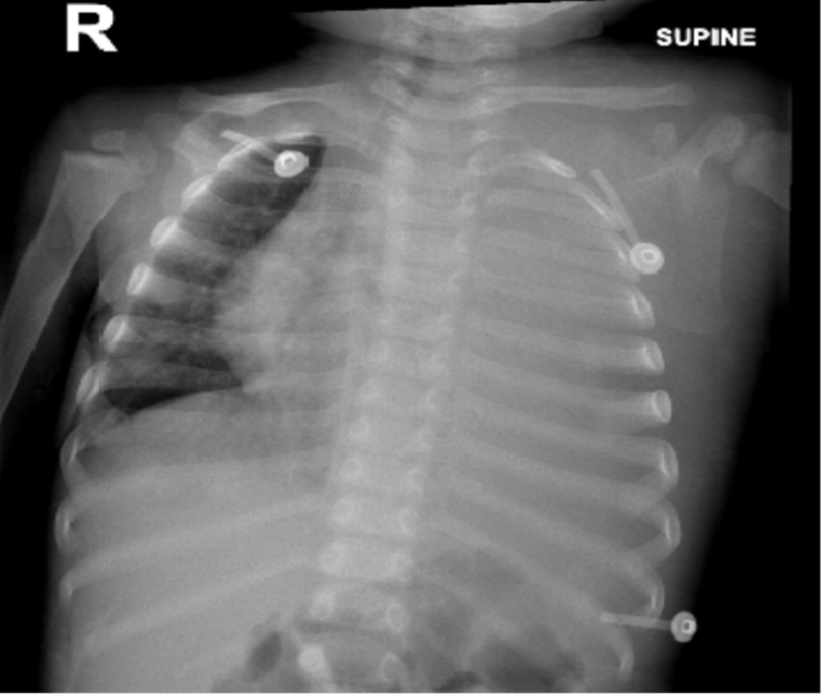
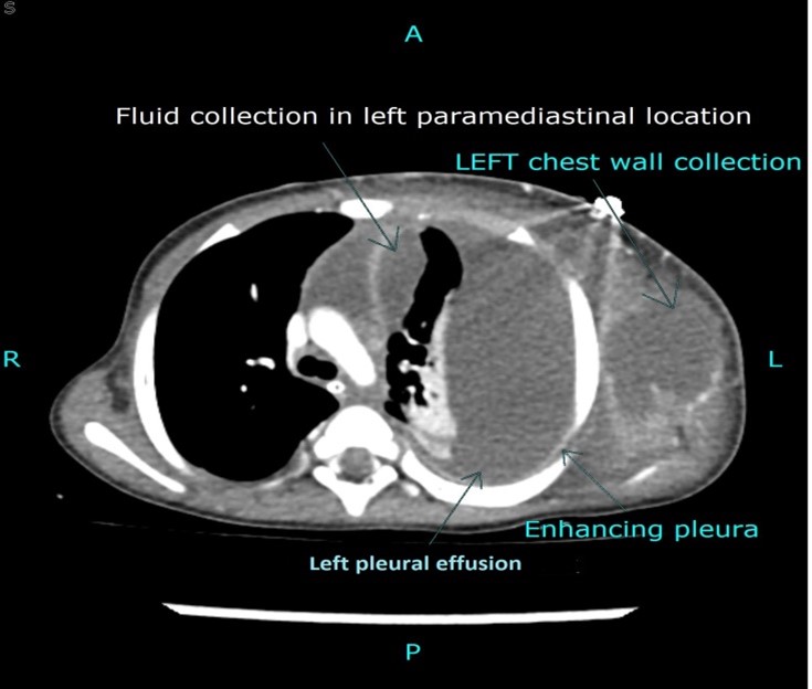
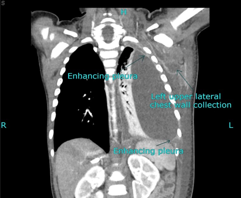
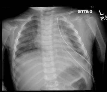
1.5-year-old previously healthy patient was referred from tertiary care to us with complaints of fever and cough for one week and respiratory distress for two days. The patient was initially managed by a general physician with supportive treatment, but due to worsening of symptoms, the patient visited another tertiary care hospital where she was admitted; a baseline workup was performed, and oxygen was provided with a noninvasive high-low nasal cannula along with broad-spectrum antibiotics. The workup revealed pleural effusion; therefore, the patient was referred to us for further management, which was consistent with respiratory distress with swelling and erythema on the left side of the chest wall extending inferiorly from the xiphoid process to the midclavicular line and superiorly up to the subclavicular region with decreased air entry on the left side of the chest. Laboratory workup revealed leukocytosis with white blood count of 58.9k/µL with a left shift 74% neutrophils, 0.1% eosinophil’s, 6.1% monocytes, 19.7% lymphocytes, hemoglobin 9.9 g/dL, platelet count 703 K/µL, C-reactive protein 150.94 mg/L, procalcitonin 4.01 ng/mL and blood gas with pH 7.47, partial pressure of carbon dioxide 34.80 mm Hg, partial pressure of oxy-gen 185 mm Hg, bicarbonate 24.7meq/L and base excess 1.5 meq/L. Chest X-ray findings were consistent with complete left hemithorax whiteout with a contralateral mediastinal shift, as obvious in fig1. Computed tomography of the chest was performed, consistent with gross left-sided pleural effusion with an enhanced pleural margin suggestive of empyema, large collection with an enhancing margin in the left lateral chest wall, and smaller fluid collection in the paramediastinal location consistent with Empyema Necessitans (EN), as shown in figure 1b & 1c. The patient was admitted to the pediatric intensive care unit, intubated, and continued on mechanical ventilation for increased work of breathing. The broad-spectrum antibiotics ceftriaxone and vancomycin were continued. The cardiothoracic surgery team was taken onboard, and left posterolateral thoracotomy and decortication were performed under general anesthesia with more than 500 ml purulent fluid drained and two chest tubes placed. The post-procedure patient went into SIRS, which was managed with inotropes, fluid resuscitation, and adjustment of ventilatory support. Cultures revealed MRSA. Pleural fluid for the AFB smear and gene-xpert were negative. An echocardiogram, which was negative for vegetation, was performed. IV ceftriaxone was discontinued, and vancomycin was switched to intravenous linezolid. Post-procedure radiography showed resolution of the pleural effusion, as shown in fig 1d and 1e. Chest tubes were removed on 7th postoperative day. The patient was treated with one week of IV antibiotics (4 days of ceftriaxone and vancomycin and 3 days IV linezolid) followed by oral linezolid for 4 weeks. The patient’s hospital course remained uneventful, and Patient was discharged in stable condition on oral antibiotics on postoperative day 9.

Figure 1a: Complete left hemi thorax white out with mediastinal shift suggestive of pleural effusion.

Figure 1b: Axial contrast enhanced CT chest showed gross left sided pleural effusion with enhancing pleural margin, suggesting empyema, laterally there is large collection with enhancing margin in left lateral chest wall. Medially, a smaller fluid collection is seen in paramediastinal location.

Figure 1c: Coronal Contrast enhanced CT image which showed gross left sided pleural effusion with enhancing pleural margin. There is large collection with enhancing margin in left lateral chest wall.

Figure 1d: Chest X ray AP view showed tracheal and mediastinal structure in normal position. Two chest tubes in placed one in apical and one in basal area. Resolution of pleural effusion in left side.

Figure 1e: Interval removal of chest tube on left sided.
Discussion
Empyema Necessitans (EN) is the extravasation of empyema from the pleural cavity into the surrounding structure, involving the chest wall. It is one of the rare complications of complicated pneumonia, and the most common organisms that are usually associated with EN are Mycobacterium tuberculosis and Actinomyces israelli, whereas other rare organisms include Staphylococcus aureus, as in our case, Pseudomonas, and Streptococcus.
The diagnostic study of choice is contrast-enhanced chest CT, which shows the extension of fluids and involvement of the underlying structure [3,4]. Delay in diagnosis can lead to morbidity and mortality and involvement of other mediastinal structures, including the abdominal wall, paravertebral space, vertebrae, esophagus, bronchus, mediastinum, diaphragm, pericardium, flank, breast, and retroperitoneum [5]. Treatment guidelines have not yet been established due to the limited literature available on this disease, but treatment options include both surgical drainage with decortication and antibiotic administration [6]. Further studies are required to determine the duration of antibiotic use in EN. As in our case report, we also managed empyema necessitans with surgical drainage, decortication, and antimicrobial administration. The first case of EN associated with MRSA was described by Stall et al(2005) in an 8 months old who was treated with simple chest tube placement and a total of 21 days of antibiotic treatment, which included 10 days of intravenous vancomycin [7]. Another case of EN associated with MRSA was reported by Moore et al (2006) in 3 months old infant who was treated by surgical drainage and tube thoracotomy and decortication with a total of 21days of antibiotic treatment [8]. Preston reported 5-year-old child with MRSA who was treated with ultrasound-guided chest tube placement and 21 days of antibiotic therapy [9]. Compared to the above-mentioned case reports, we prescribed antibiotics for a total 4 weeks of duration to avoid relapse.
Conclusion
Empyema necessitans is a rare complication of pneumonia. Any child who presents in the emergency department with respiratory symptoms, chest wall mass, and empyema necessitans should be considered as a differential diagnosis. Successful treatment options include surgical drainage with decortication, and a prolonged antibiotic course.
Disclosures:
Ethical approval and consent to participate: Informed Verbal consent was obtained from the parents of the patient for the case details to be used for any publication.
Consent to publish: Written informed consent to publish was obtained from the parents of the patient for publication of this case report in a journal as well for other study purposes.
Availability of data and material: Case details are not publicly available because the data is patient medical records but are available from the corresponding author upon reasonable request
Author’s contribution: RO, YT and AM were involved in the literature search of a topic, AR: Data Acquiring, Writing the manuscript and final revision of manuscript WA: final revision of manuscript NR: Concept and design, interpretation of data, final revision of manuscript. All authors have read and approved final manuscript.
Conflicts of interest: None
Acknowledgments: None
Funding: None
References
- Kono SA, Nauser TD. Contemporary empyema necessitatis. . The American journal of medicine, 2007; 120: 303-305.
- Akgül AG, Örki A, Örki T, et Approach to empyema necessitatis. World J Surg, 2011; 35: 981-984.
- Gupta DK, Sharma Management of empyema-Role of a surgeon. J Indian Assoc Pediatr Surg, 2005; 10: 3.
- Chan W, Keyser-Gauvin E, Davis GM, Nguyen LT, Laberge Empyema thoracis in children: a 26-year review of the Montreal Children's Hospital experience. J Pediatr Surg, 1997; 32: 870-872.
- Reyes CV. Cutaneous tumefaction in empyema necessitatis. Int J Dermatol. 2007; 46: 1294-1297.
- Edaigbini SA, Anumenechi N, Odigie VI, Khalid L, Ibrahim AD. Open drainage for chronic empyema thoracis; clarifying misconceptions by report of two cases and review of Archives of International Surgery, 2013; 3: 161.
- Stall worth J, Mack E, Ozimek C. Methicillin-resistant Staphylococcus aureus empyema necessitatis in an eight-month-old South Med, 2005; 98: 1130-1131.
- Moore FO, Berne JD, McGovern TM, et Empyema necessitatis in an infant: a rare surgical disease. J Pediatr Surg, 2006; 41: 5-7.
- Pugh CP. Empyema Necessitans a Rare Complication of Methicillin-Resistant Staphylococcus Aureus Empyema in a Pediatr Infect Dis J, 2020; 39: 256-257.

