CT Scan and Obturator Hernia
Salah Ben Elhend1,*, B Ait Idir2 and A El Kharras1
1Department of Radiology, 1st Medical and Surgical Military Center, Agadir, Morocco
2Department of Surgery, 1st Medical and Surgical Military Center, Agadir, Morocco
Received Date: 09/02/2023; Published Date: 25/04/2023
*Corresponding author: Salah Ben Elhend, Department of Radiology, 1st Medical and Surgical Military Center, Agadir, Morocco
Abstract
Bowel obstruction by obturator hernia is a rare surgery and imaging condition. Caused by incarcerated bowel segment in obturator foramen, it occurs generally among elderly women. There is no specifics clinical finding and most often, the hernia is not detected in physical examination. We report the case of mechanical bowel obstruction due to incarcerated obturator hernia in elderly thin woman. Computed Tomography
(CT scan) showed an incarcerated bowel with a fatty mass in the left obturator foramen.
Generally, CT scan showed incarcerated bowel with hydrometric level. Intravenous contrast and a neutral luminal contrast improved visibility of bowel wall enhancement and thickness. Bowel wall necrosis and perforation radiological signs are rarely seen, and must be carefully researched: pneumatosis intestinalis, pneumatosis portalis pneumoperitoneum, submucosal hemorrhage and variable amounts of free fluid.
CT scan, is recommended for early diagnosis, to avoid complications and for a surgical cure in better conditions.
Keywords: Hernia; Obturator foramina; CT scan
Introduction
Obturator hernia is a bowel, generally small bowl, passing in the obturator foramina, frequently observed in elderly women, with emaciation and chronical disease increasing pressure in abdominal cavity. Its uncommon occurring in 1% of population, and can lead to the usual complications of a strangled hernia, such as acute bowel obstruction and necrosis.
Case Report
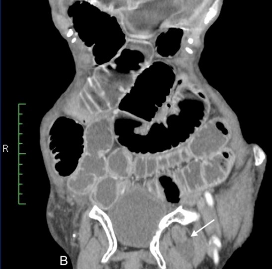
We describe a case of an 80-year-old lady, with no history of chronical disease or previous abdominal surgery. Her family reported a significant weight loss over the last year. She was admitted to the emergency department for abdominal distension, obstipation and bilious vomiting for 07 days. Clinical examination found a thin patient with diffuse abdominal pain, central distension of abdomen with generalized tenderness. Her full blood count, renal function, and liver function tests were normal. An abdominal X-ray showed dilated loops of small bowel. A Computer Tomography (CT scan) revealed an incarcerated left obturator hernia (Figure 1). After this unexpected CT finding, a meticulous neurological examination found inner thigh pain that may extend to the knee on internal rotation of the hip secondary to the irritation o the left obturator nerve, masked on admission by diffuse abdominal pain. The left obturator hernia defect was closed by suturing edges of obturator orifice, reinforced by polypropylene mesh. The postoperative period in the hospital was 02 days, and passed uneventfully.

Figure 1: Hypochromic macules. pityriasis versicolor like on the trunk.
Discussion
Reported incidence of obturator hernia ranges from 0.05% to 1.4% of all hernias and are responsible for 0.2% to 1.6% of intestinal obstructions [1]. There have only been 400 published cases, noted in a literature review from 1966 to 2000 [2].
Despite the Romberg-How ship, sign described with great sensitivity, symptoms and clinical findings are usually non-specific, for that, obturator hernia is difficult to diagnose and rarely diagnosed before CT scan. Our patient presented with complaints of acute intestinal obstruction, with no past history of recurrent attacks of intestinal obstruction.
Computed Tomography scan, is recommended for early diagnosis. Some authors recommend laparotomy to diagnose and treat at the same time, whereas others prefer preoperative noninvasive diagnostic methods such as CT scan [3,4].
Generally, CT scan showed incarcerated bowel in the obturator foramen with hydrometric level (Figure 2,3). Intravenous contrast and a neutral luminal contrast improved visibility of bowel wall enhancement and thickness. Bowel wall necrosis and perforation radiological signs are rarely seen, and must be carefully researched: pneumatosis intestinalis, pneumatosis portals pneumoperitoneum, submucosal hemorrhage and variable amounts of free fluid [5].
Our case was interesting, showing an obturator hernia in the left side, contrasting with literature data suggesting more right-side location. Other remarkable particularity of our case, was that the incarcerated bowl contained an outer edge lipoma (Figure 4), showed as a fatty mass with low density, and authors suggest increased risk of obturator herniation in presence of bowel masses.
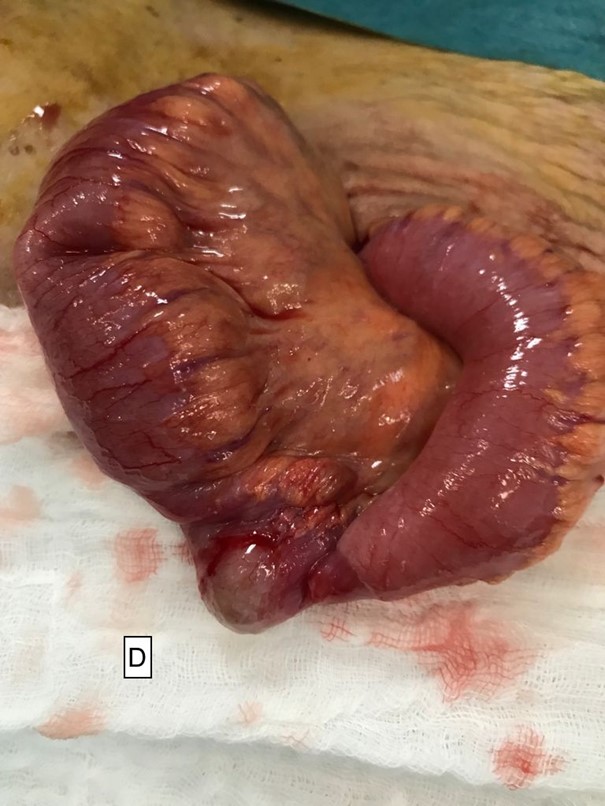
Patient was managed by laparotomy. After left femoral incision, we discover the incarcerated hernia between dilated and normal bowel (Figure 5). We try to externalize the incarcerated bowel by gentle traction. The small bowel was not necrotic, and no bowel resection was performed. Obturator orifice was repaired by suturing of the orifice edges, respecting vasculonervous pedicle, with reinforcement with polypropylene mesh: by first exposing peritoneum, fixing the polypropylene mesh, then covering it with the peritoneum.

Figure 2: Erythematous scaly plaques histologically corresponding to Bowen's disease.

Figure 2: Erythematous scaly plaques histologically corresponding to Bowen's disease.

Figure 2: Erythematous scaly plaques histologically corresponding to Bowen's disease.

Figure 3: Clinical and dermoscopic appearance of wart on the back of the foot.
Conclusion
Obturator hernia is a rare diagnostic challenging cause of acute intestinal occlusion, particularly seen in elderly emaciated women. CT scan and laparotomy, or laparoscopy if the patient's condition allows it, can decrease risk of bowel necrosis and lower morbidity and mortality.
Conflicts of interest: The authors report no conflicts of interest.
References
- Rodriguez-Hermosa JI, Codina-Cazador A, Maroto-Genover A, Puig- Alcantara J, Sirvent-Calvera JM, Garsot-Savall E, et al. Obturator hernia: clinical analysis of 16 cases and algorithm for its diagnosis and treatment. Hernia, 2008; 12: 289-297.
- Losanoff JE, Richman BW, Jones JW. Obturator hernia. J Am Coll Surg, 2002; 194: 657-663.
- Pélissier E, Ngo P, Armstrong O. Traitement chirurgical des hernies obturatrices. EMC (Elsevier Masson SAS, Paris), Techniques chirurgicales - Appareil digestif, 2010; 40-155.
- Nishina M, Fujii C, Ogino R, et al. Preoperative diagnosis of obtu- rator hernia by computed tomography in six patients. Emerg. Med, 2001; 20: 277–280.
- Furukawa A, Kanasaki S, Kono N, et-al. CT diagnosis of acute mesenteric ischemia from various causes. AJR Am J Roentgenol, 2009; 192 (2): 408-416.

