Unusual Liver Hydatid Cyst Fistulization
Choayb S*, Harras El Y, Allali N, Chat L And Haddad El S
Children’s Hospital, Radiology Department, Mohamed V University, Rabat, Morocco
Received Date: 30/01/2023; Published Date: 08/03/2023
*Corresponding author: Safaa Choayb, Children’s Hospital, Radiology Department, Mohamed V University, Rabat, Morocco
Abstract
The fistulization of a hydatid cyst in the gallbladder is very rare. We present a case of an 11-year-old boy presenting abdominal pain and tenderness of the right upper quadrant. Imaging revealed a liver hydatid cyst communicating with the gallbladder.
Introduction
Hydatid disease also known as hydatidosis is a major health problem in endemic areas, especially the Mediterranean countries. Liver is the most typical location. It can be complicated by a rupture in the hepatic biliary system. The communication between the hydatid cyst and the gallbladder is rare.
Case Report
An 11-year-old boy without any past medical history consults for right hypochondrial discomfort. Medical examination revealed hepatomegaly and diffuse tenderness in the right upper quadrant. The patient did not present a fever or jaundice. Hydatid serology was positive. The other laboratory findings were normal.
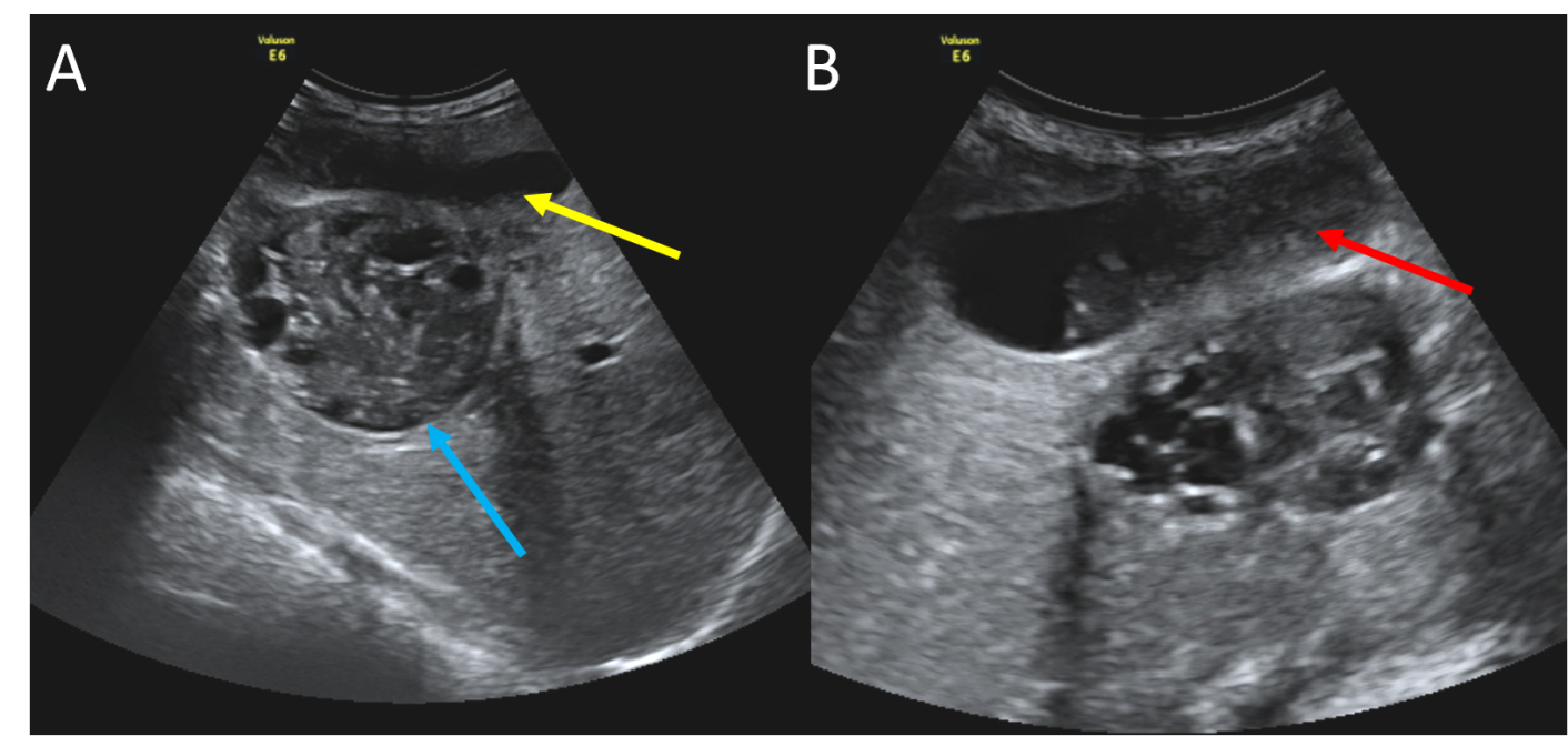
Abdominal ultrasound showed a multiloculated cystic lesion in the 5th segment of the liver communicating with the gallbladder that contained echogenic material (Figure 1). The common biliary duct was not dilated.
CT scan revealed a deformed liver hydatid cyst communicating with the gallbladder via a defect in its wall (Figure 2).
The patient underwent surgery. The exploration confirmed the imaging findings. The postoperative period was uneventful.

Figure 1: (A and B) Transverse sonograms of the liver showing a multivesicular hydatid cyst (blue arrow) juxtaposed to the gallbladder (yellow arrow) containing echogenic material (red arrow).

Figure 2: Axial abdominal CT scan showing the hydatid cyst (blue arrow), next to the gallbladder and the defect of its wall.
Discussion
Hydatid disease is a serious health issue in endemic locations, particularly the Mediterranean countries. The most prevalent location is the liver.
Liver hydatid cyst grows at a variable rate. The cyst causes pressure on the adjacent parenchyma and may rupture inside the biliary tree. Communication with the hepatic bile ducts is most frequent. Fistulization between the liver hepatic cyst and gallbladder is a rare event [1].
The rupture can be contained (endocyst is torn but the content is confined within the pericyst), communicating (tear of the endocyst with loss of the cyst content via small biliary ducts), or direct (tear of both endocyst and pericyst, with the spilling of the cyst content into the peritoneal cavity).
Clinically, the rupture can be occult and usually silent but may be accompanied by suppuration or evolve towards a frank rupture. In frank rupture, the daughter vesicles or ruptured membranes pass into the biliary tract, and intermittent or progressive obstructive jaundice, cholangitis, or septicemia may occur [2].
Cyst hydatid-gallbladder fistula may sometimes rupture and cause the spreading of the cyst content into the peritoneal cavity which can be complicated by peritonitis or peritoneal abscess. Cholangitis rarely occurs as the fistulous communication needs to be large enough to let the cyst material pass into the gallbladder. Also, the cystic duct should be wide and short enough to pass these materials into the bile duct [3].
Ultrasound and CT are used for radiological diagnosis by revealing direct and indirect features. A deformed cyst is indirect evidence of rupture. The existence of echogenic debris is suggestive of vesicles or membranes inside the extrahepatic biliary system. The visualization of the cyst wall defect or communication between the cyst and the biliary system is the only direct sign (only seen in 20% of the cases) [4,5].
Treatment is surgical consisting of treating the hydatid cyst, cholecystectomy, and verification of the vacuity of the bile ducts [6].
Conclusion
Rupture of liver hydatid cyst in the gallbladder is rare. It can be a life-threatening situation. Ultrasound and CT can help in establishing the diagnosis preoperatively to avoid further complications.
Conflict of Interests: The authors declare that there is no conflict of interests regarding the publication of this paper.
Author Contributions: All authors contributed equally to this work
References
- Hassan R, Fahmi Y, Khaiz D, Elhattabi K, Bensardi Fz, Berrada S, et al. Total rupture of hydatid cyst of liver in to common bile duct : a case report. Pan African Medical Journal, 2014; 19: 370.
- Avcu S, Ünal Ö, Arslan H. Case report Intrabiliary rupture of liver hydatid cyst : a case report and review of the literature. Cases Journal, 2009; 2: 6455.
- Bilgi Kırmacı M, Akay T, Özgül E, Yılmaz S. Cholecysto-Hydatid Cyst Fistula : A Rare Cause of Cholangitis. Am J Case Rep, 2020; 21: e921914.
- Aslan S. A rare cause of obstructive jaundice in adolescent patient : Ruptured hydatid cyst into the biliary duct. Annals of Medical Research, 2019; 26(11): 2712-2714.
- Chaouch MA, Mesbahi M, Ghannouchi M, Rebhi J, Sboui, Tlili Y, et al. Case Report of Uncommon Frank Fistulous Communication between Liver Hydatid Cyst and Gallbladder. JOP. J Pancreas, 2019; 20(5): 121-123.
- Ferjaoui W, Talbi G, Karouia S, Omrani S, Bayar R, Khalfallah MT. Rare Fistulous Communication Between a Hepatic Hydatid Cyst and the Gallbladder : a Case Report. Indian Journal of Surgery, 2020.

