Amnioexcel® Plus Graft and its Prospective Benefits in Salvaging a Below- the- Knee- Amputation Stump Wound
Shaniah Holder1,*, Gabrielle Unbehaun2, Ulochi Nwagwu2, Christopher Meusburger3 and Frederick Tiesenga4
1Department of Medicine, American University of Barbados School of Medicine, Barbados
2Department of Medicine, Saint George’s University School of Medicine, Grenada
3Department of Medicine, Saint James School of Medicine, Anguilla
4Department of Medicine, Department of Surgery, Community First Hospital, USA
Received Date: 30/01/2023; Published Date: 06/03/2023
*Corresponding author: Shaniah Holder, Department of Medicine, American University of Barbados School of Medicine, Barbados
Abstract
Below-the-Knee-Amputation (BKA) is a life-saving procedure and is associated with a better functional outcome in persons with severe vascular comorbidities when compared to Above-the-Knee Amputation (AKA). Amputation stump wounds are associated with pain and an increased risk of undergoing a subsequent AKA, which adversely affects the quality of life. The AmnioExcel® Plus (Integra, Princeton NJ) allograft derived from placental tissue can be used as a revolutionary tool in the management of complicated amputated wounds and the possible benefit of salvaging limbs. Amniotic cells possess stem-cell-like proliferation and differentiation properties, making them great sources for wound healing. We present a case of a post-BKA patient with a nonhealing stump wound, who was subsequently managed with AmnioExcel® Plus and wound vacuum placement. This report aims to examine the properties, indications, and advantages of implementing this graft as a promising treatment for transtibial amputation wounds and the prevention of subsequent amputations.
Keywords: AmnioExcel; Diabetes Mellitus; Nonhealing ulcer; BKA stump; Tissue regeneration; Amputations; Allograft tissue
Introduction
Diabetes Mellitus (DM) and Peripheral Vascular Disease (PVD) affect over 37 million Americans and are the two leading causes of impaired wound healing [1]. Peripheral neuropathy is one of the most common complications arising from diabetes and PVD and has an estimated prevalence of 28% [2]. This precipitates the formation of nonhealing ulcers which are often preceded by mild trauma [2]. Roughly 150,000 patients undergo lower extremity amputations annually, and diabetic neuropathy remains a principal etiology [3]. Therefore, suitable management is required to prevent further complications and lifelong disabilities.
AmnioExcel® Plus (Integra, Princeton NJ) is a dehydrated trilaminar allograft derived from placental tissue [4]. It consists of multiple molecular factors resulting in scar attenuation, angiogenesis, inflammatory cell suppression, and protein preservation [4]. Crucially, it does not express human leukocyte antigens, which inherently reduces immunogenicity and rejection risk [5]. This has transformative potential for accelerating wound repair when compared and added to conventional regimens for instance, negative pressure wound therapy (NPWT) with the use of a vacuum-assisted closure (VAC) device is a commonly implemented modality for both acute and chronic wounds [6]. These devices became FDA-approved in 1995 and have been used by millions since they induce localized hypoxic tissue states, stimulate angiogenesis, and promote wound resolution [6]. The use of AmnioExcel® Plus with wound vac placement may enhance the wound-healing process and potentially benefit patients with nonhealing wounds caused by the aforementioned health morbidities [4]. The benefits and extent of healing support, tissue restoration, and improved health outcomes in otherwise non-viable amputation stump wounds require more comprehensive research.
We present a patient with a nonhealing diabetic ulcer on a previously performed below-the-knee amputation stump site. He was an excellent candidate for AmnioExcel® Plus allografting to salvage his limb and prevent an additional amputation. Five days after initial placement with accompanied wound vac placement, enriched areas of newly formed granulation tissue had noticeably begun to develop with increased tensile strength and cellular integration.
Case Presentation
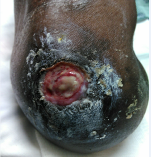
We present a 63-year-old male with a past medical history significant for DM 2, PVD, hypertension, and substance use disorder and a surgical history remarkable for a right BKA of performed two years ago due to his comorbidities. He presented to the Emergency Department (ED) with a chronic nonhealing stump wound. The patient stated that he just “bumped” the stump a couple of months after the procedure, and a wound formed, which never healed. On physical exam, the leg was mildly edematous with a wound that was 2.9 cm in length, 1.9 cm in width, and 0.3 cm in depth. The wound bed was covered with 100% wet, yellow slough with a moderate amount of faint-smelling seropurulent exudate. The edges appeared to be well-defined with a dry, intact peri wound. The patient endorsed pain which was exacerbated by the assessment. (Figure 1) below shows the infected stump when the patient arrived at the ED.

Figure 1: Infected BKA stump wound with purulent drainage and erythema visualized.
These findings were concerning for cellulitis and possible osteomyelitis, which warranted further investigation with imaging. The patient was placed on prophylactic piperacillin-tazobactam and Computed Tomography (CT) of the right lower extremity with intravenous contrast was conducted. Results showed subcutaneous edema consistent with cellulitis and a central lytic area involving the metaphyseal region of the distal right femur suspicious for chronic osteomyelitis. (Figure 2) below highlights the CT findings of osteomyelitis of the femur.

Figure 2: Central lytic area involving the metaphyseal region of the distal right femur indicating possible chronic osteomyelitis.
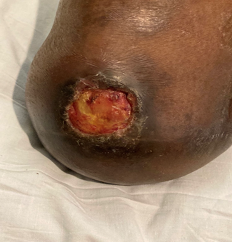
The patient was diagnosed with an infected stump wound indicating debridement of the tissue in addition to prophylactic antibiotic use. However, due to the risk of poor wound healing after the removal of the infected tissue, it was decided that cell graft placement was necessary to salvage the stump and prevent further amputations in the future. The patient underwent a right below-knee amputation wound debridement with AmnioExcel® Plus placement. All necrotic skin and subcutaneous tissue and debris were sharply excised, the bone was not exposed and an AmnioExcel® Plus graft was placed, secured, and wrapped in sterile dressing. The patient had no postoperative complications and two days later the dressing was changed with wound vac placement to optimize and accelerate the healing process. Eight days postoperative, there was visible integration of the cell within the wound and increased granulation tissue formation. The new wound measurements were 2.1cm in length, 2.5cm in width, and 0.2cm in depth indicating cell regeneration. Figure 3 below shows the BKA stump wound eight days after the placement of AmnioExcel® Plus.

Figure 3: Image showing cell integration and increased granulation tissue formation on post-op day 8.
The surgical team signed off, and the patient was instructed to follow up with wound care.
Discussion
Delayed wound healing is a long-known consequence of poorly controlled DM and PVD and is the leading cause of lower extremity amputation and subsequently increased mortality.
Chronic wounds provide a challenge to physicians because these wounds fail to progress through the appropriate stages of healing [7]. These wounds often have excessive proinflammatory cytokines, extracellular matrix destruction, and prolonged infection occurrence; as well as the deficiency of functional stem cells, collagen, and angiopathic factors [7]. Historically, the management of these wounds focused predominantly on infection prevention, while more recently, there is a greater emphasis on providing active support to the wound healing process. VAC devices have greatly improved the rate of healing in these challenging wounds by increasing the rate of granulation tissue development and vascularity of these wounds, while also decreasing bacterial growth [6]. However, these machines can be cumbersome to patients and they lead to slower ambulation, therefore research should continue to investigate adjunctive treatments for these wounds [8]. The use of skin substitutes in these amputation site wounds provides an opportunity to ameliorate some of these challenges and recent literature has indicated that amniotic membrane grafts have an efficacy of as high as 86% to 90% in healing difficult wounds [5].
Skin substitutes are often bilaminar with an amnion and chorion component or unilaminar with only an amnion component [9]. The AmnioExcel® Plus is a trilaminar (amnion-chorion-amnion) allograft derived from donated human placental tissues. These placentas must meet the requirements of the rigorous screening and testing process before sterilization and dehydration [4]. The extra amnion layer provides additional support and reduces graft degradation [4,9]. These grafts are suggested to be non-immunogenic due to the innate immunologically privileged features of the placenta [10,11]. It reduces the expression of the Major Histocompatibility (MHC) protein complex, and the expression of apoptotic proteins, and stimulates the release of immunosuppressive and immunomodulatory molecules [10-12].
The amnion components provide the majority of the structural support for the graft and the wound with extracellular matrix proteins, while the chorion component provides ample type I and III collagen, laminin, fibronectin, and signaling molecules [9]. The amniotic membrane induces cell migration and proliferation with increased levels of transforming growth factor β and vascular endothelial growth factor [11]. The addition of these molecules to the wound site has been shown to greatly increase the rate of epithelialization, and angiogenesis, while decreasing inflammation and increasing the overall probability of successful wound closure [11,13,14]. In addition, the release of anti-inflammatory cytokines and IL-10 has an analgesic effect [5]. The graft also provides physical coverage and protection to the regenerating nerve twigs which further helps in pain alleviation [5].
Placement of an AmnioExcel® Plus graft is recommended in the immediate postoperative management of a BKA site. In addition to cell placement, sterile dressing with a knee immobilizer accompanied by serial examinations for signs of wound dehiscence, infection, or bleeding should be implemented [15]. AmnioExcel® Plus use is indicated as a wound-healing support for acute below-knee amputation wounds or as an adjunctive treatment to VAC devices to salvage below-knee amputation sites, especially in those with complex comorbidities such as diabetes mellitus or PVD [4]. Some contraindications to AmnioExcel® Plus graft use are patients with an allergy or sensitivity to ethanol due to trace amounts of ethanol potentially present due to the preparation process [4]. In addition, placental allografting is contraindicated in any patient with active infection [4].
The AmnioExcel ® Plus graft is a promising therapeutic option when salvaging BKA site wounds. As seen with our patient, these grafts can increase epithelization, granulation tissue development, and angiogenesis while decreasing inflammation and pain. In addition, these grafts have minimal contraindications and complications, providing a safe way to encourage timely healing of these chronic wounds and reducing the morbidity of these patients.
Conclusion
In this report, the AmnioExcel® Plus placental allograft membrane showed promising granulation results, tissue reepithelialization, and reduced pain in the patient. These placental membranes are transformational as it offers targeted wound healing due to their biological properties, immunologic characteristics, and analgesic effects. As with this patient, diabetes mellitus and vascular disease impair the multiple stages of wound healing. This leads to hard-to-treat infections, amputations, and frequent hospitalizations. AmnioExcel® Plus placental allograft is groundbreaking compared to standard wound care, as the trilaminar structure promotes paracrine effects for reduced healing time and tissue regeneration. Although the results were reassuring, future research into placental allografts and their possibility of salvaging BKA stumps from further amputation is necessary.
Author Contributions:
Shaniah Holder: Concept and Design of study, Acquisition of data, drafting article, revising article. Guarantor Author
Gabrielle Unbehaun: Concept and Design of study, drafting article intellectual content, revising article
Ulochi Nwagwu: Acquisition of data, drafting article, revising article
Christopher Meusburger: Acquisition of data, drafting article, revising article
Frederick Tiesenga: Final Approval, Intellectual Content, revising article
Informed Consent: A signed statement of informed consent was obtained for publication of the details of this case
Competing Interests: None
Grant Information: None
References
- Type 2 Diabetes, 2021.
- Hicks CW, Selvin E. Epidemiology of Peripheral Neuropathy and Lower Extremity Disease in Diabetes. Curr Diab Rep, 2019; 10: 86. DOI: 10.1007/s11892-019-1212-8.
- Lower Extremity Amputation, 2022.
- A Prospective, Multi-center, randomized, parallel-group study comparing AmnioExcel® Plus Placental Allograft Membrane to Apligraf® Bi-layered Skin Substitute and Standard of Care Procedures in the Management of Diabetic Foot Ulcers, 2019.
- Schmiedova I, Dembickaja A, Kiselakova L, et al. Using of Amniotic Membrane Derivatives for the Treatment of Chronic Wounds. Membranes (Basel). 2021; 11: 941. DOI: 10.3390/membranes11120941.
- Agarwal P, Kukrele R, Sharma D. Vacuum Assisted Closure (VAC)/Negative Pressure Wound Therapy (NPWT) for difficult wounds: A review. J Clin Orthop Trauma, 2019; 10(5): 845-848. DOI: 10.1016/j.jcot.2019.06.015.
- Frykberg RG, Banks J. Challenges in the Treatment of Chronic Wounds. Adv Wound Care (New Rochelle). 2015; 4: 560-582. DOI: 10.1089/wound.2015.0635.
- Sim Chap H, Vickneswaren L, Regidor III D. Vacuum-Assisted Wound Dressing For Shorter Wound Healing Time: A Meta -Analysis. MJMR, 2022. DOI: 10.31674/mjmr.2022.v06i04.004.
- Bonvallet PP, Damaraju SM, Modi HN, et al. Biophysical Characterization of a Novel Tri-Layer Placental Allograft Membrane. Advances in Wound Care, 2022; 11: 43-55. DOI: 1089/wound.2020.1315.
- Roy A, Mantay M, Brannan C, et al. Placental Tissues as Biomaterials in Regenerative Medicine. Biomed Res Int, 2022; 67: 514-556. DOI: 10.1155/2022/6751456.
- Pogozhykh O, Prokopyuk V, Figueiredo C, et al. Placenta and Placental Derivatives in Regenerative Therapies: Experimental Studies, History, and Prospects. Stem Cells Int, 2018; 4: 837-930. DOI: 10.1155/2018/4837930.
- Riley JK. Trophoblast immune receptors in maternal-fetal tolerance. Immunol Invest, 2008; 37: 395-426. DOI: 10.1080/08820130802206066.
- Lakmal K, Basnayake O, Hettiarachchi D. Systematic review on the rational use of amniotic membrane allografts in diabetic foot ulcer treatment. BMC Surg. 2021; 1: 87. DOI: 10.1186/s12893-021-01084-8.
- Snyder RJ, Shimozaki K, Tallis A, et al.: A Prospective, Randomized, Multicenter, Controlled Evaluation of the Use of Dehydrated Amniotic Membrane Allograft Compared to Standard of Care for the Closure of Chronic Diabetic Foot Ulcer. Wounds, 2016; 3: 70-77. PMID: 26978860.
- Below Knee Amputation, 2022.

