Ectopic Left Coronary Artery Connected to the Right Coronary Artery
Otman Bouzouba*, Amal Hsain and Abdoulwahab Karimou Bondabou
Department of Cardiology, Ibn Sina Hospital, Rabat, Morocco
Received Date: 27/12/2022; Published Date: 20/01/2023
*Corresponding author: Otman Bouzouba, Resident, Department of Cardiology, Ibn Sina Hospital, Rabat, Morocco
Abstract
The ectopic coronary artery connected to the contralateral artery is part of the anomalies of proximal connection of the coronary arteries (ANOCOR) which are rare in adults without structural congenital heart disease. It is characterized by the presence of a single coronary ostium with an initial ectopic course. Its prognosis depends mainly on the type of the ectopic course in relation to the adjacent cardiac structures. We report the case of a 62-year-old patient with a fortuitous discovery of an ectopic left coronary artery connected to the right coronary artery with an initial retro-aortic course, an anatomical shape with a good prognosis in general. The patient was pauci symptomatic accusing isolated atypical precordialgia for 2 years. The electrocardiogram does not show repolarization disorders. The chest X-ray shows a normal cardiac silhouette with no signs of overload. Echocardiography shows normal sized heart chambers with preserved LV systolic and diastolic function. The stress test was contentious. Coronary angiography made it possible to suspect the diagnosis in view of the impossibility of intubation of the left coronary artery and the demonstration of a single right coronary ostium with an ectopic left coronary connected to the proximal right coronary artery. The diagnosis was confirmed by CT scan showing an ectopic left coronary artery connected to the right coronary artery with an initial retro-aortic course.
Keywords: Coronary abnormalities; Coronary ostium; Ectopic coronary artery
Introduction
The ectopic coronary artery connected to the contralateral artery is a rare anomaly of proximal connection of the coronary arteries. Its main differential diagnosis is the single coronary artery which is also characterized by the existence of a single ostium and the absence of an initial intramural aortic course, but is differentiated from the ectopic coronary artery by the absence of an initial ectopic course.
We report the case of a 62-year-old woman in whom an ectopic left coronary artery connected to the right coronary was discovered by chance.
Case Presentation
-This is Mrs. K.H, 62 years old, married and mother of 2 children.
-She has cardiovascular risk factors, apart from menopause dating back 18 years, hypertension evolving for 15 years under well-balanced dual therapy, dyslipidemia for more than 10 years under lipid-lowering diet alone, and a history of hospitalization for 3 days for arrhythmia due to atrial fibrillation.
-Patient admitted to our service for palpitations with sudden beginnings and ends associated with precordial chest pain, moderate, brief, less than 3 min, radiating to the back, without notion of faintness or syncope or sensorimotor deficit.
-The clinical examination was strictly normal and found a patient in good general condition, hemodynamically and respiratory stable.
-The biological assessment was normal.
-The electrocardiogram is in regular sinus rhythm with a normal axis of the heart, without visible repolarization disorders.
-An echocardiography was performed and showed a normal size of LV with non-hypertrophied walls, good systolic function with an LVEF of 68%, normal filling pressure, dilated left atrium, normal function of the right heart.
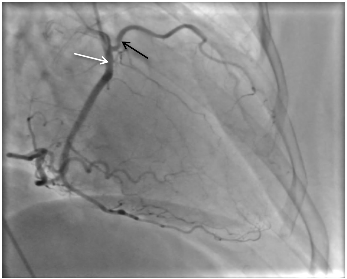
-Facing this atypical precordialgia, a coronary angiography was performed and allowed to suspect an abnormal connection of the left coronary artery due to the impossibility of intubating the left common trunk: the left coronary artery is connected to the proximal part of the right coronary artery without a visible lesion (Figure 1).
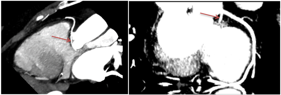
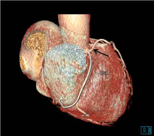
-On these data, a COROSCANNER was indicated to better explore the congenital anomaly of the coronary arteries and identify its course. It objectified an abnormal connection of the left main trunk to the proximal part of the right coronary artery then a retro aortic path without an intra parietal aortic path, bypassing the anterior surface of the aortic root to join its usual path (Figure 2-6). The coronary arteries are free from stenosis.
- The patient was put under medical treatment with therapeutic abstention for her ANOCOR.

Figure 1: Angiographic image showing an abnormal connection of the left main trunk (black arrow) with the right coronary artery (white arrow) (left anterior oblique view).

Figure 2: CT scan image: Left coronary artery connection abnormality: Left common trunk (Arrow) connected to the proximal part of the right coronary artery with a single right coronary ostium.

Figure 3: 3D reconstruction: Abnormal connection of the left coronary artery (Arrow) to the proximal part of the right coronary artery with the presence of a single coronary ostium on the right. RCA= right coronary artery.

Figure 4: CT scan image: Single right coronary ostium (Arrow).
Figure 5: CT scan image: Retro-aortic trajectory of the left common trunk.
RCA: Right coronary artery; LCT: Left common trunk
Figure 6: CT scan image: the left common trunk returns to its normal path and divides into the anterior interventricular and the circumflex arteries.
CA: Circumflex artery; LAD: Left anterior descending artery
Discussion
The embryogenesis of the coronary vasculature begins with sinusoids located in the interventricular and atrioventricular sulci. The connection of the coronary arteries to the aorta occurs after the development of intracardiac septa and arterial trunks (aorta and trunk of the pulmonary artery). There is first a separation of the aortic and pulmonary rings (initially joined) with formation of the sub-pulmonary septum which distances the developing coronary arteries, and of the sub-aortic septum which attracts them. Thus, the connection of the coronaries to the aorta is made by penetration into the right anterior and left anterior aortic sinuses, also called coronary sinuses. This is why we prefer to use the term anomaly of coronary connection rather than coronary birth [1]. It is therefore concluded that the position of the arterial trunks and their relationship to each other play an important role in the connection of the coronary arteries. In congenital heart disease with malformation of the arterial trunks, ANOCORs are frequent [2], whereas in the case of our patient, in the absence of structural congenital heart disease, the precise mechanism of the abnormal connection of the coronary artery remains unknown.
The ectopic coronary artery connected to the contralateral artery is defined by the existence of a single coronary ostium connected in the usual sinus with a non-ectopic coronary artery following its usual course at the level of the cardiac structures, and an ectopic artery (coronary left or right coronary) generally connected to the first millimeters of the contralateral artery (Figure 7) or one of its branches. We come to understand that the ectopic coronary artery necessarily presents an abnormal initial course (distance between the connection with the non-ectopic artery and the area where the ectopic artery joins its usual myocardial territory) of variable length related to the arterial trunks and to the adjacent cardiac structures allowing it to reach its myocardial territory which is irrigated by the anterograde pathway. There are four possible pathways (Figure 8) which are: a) a pre-infundibular course, b) a retro-infundibular course which goes along the pulmonary infundibulum and the sub-aortic septum, c) a pre-aortic course, or d ) a retro aortic course.
The course of the ectopic artery is juxta mural to the aorta (very close to the aorta but without a common media) in the case of a pre-aortic or retro-aortic course, and could not theoretically be intra-mural (with a common media to the aorta and to the ectopic artery) in the case of an ectopic coronary artery connected to the contralateral artery.
This is a rare congenital anomaly but whose angiographic prevalence remains difficult to determine due to the scarcity of studies that distinguish ectopic connections with the contralateral artery from the single coronary artery. The only study that clearly differentiates them finds a prevalence of 0.046% for ectopic coronary artery with a higher frequency of left ectopic coronary artery than right ectopic coronary artery [3].
The coronary angiography generally makes it easy to diagnose an ectopic coronary artery connected to the contralateral artery, but still does not give good precision on the initial ectopic course with a risk of interpretation errors or even confusion, especially for the distinction between a pre-aortic course and a retro-infundibular course.
It is the cross-sectional imaging, and more particularly the CT scan, which has become the gold standard in this field for the diagnosis and the precision of the initial course, especially to distinguish a pre-aortic course from a retro infundibular course. During a pre-aortic course, the ectopic artery will pass very close to the usual connection of the absent ostium, whereas during a retro-infundibular course, the ectopic artery remains at a distance from the usual connection of the absent ostium (Figure 8).
This connection anomaly may be associated with a risk of sudden death on exertion, especially in high-level athletes [4]. This risk is dependent, according to retrospective studies, on the type of initial ectopic trajectory and concerns only the pre-aortic trajectory [5], and therefore the pre-infundibular, retro-infundibular or retro-aortic trajectories are not considered to be at risk. This agrees with the case of our patient who is pauci symptomatic without a personal history of resuscitated sudden death.
Sudden death is probably related to ventricular fibrillation linked to the existence of areas of fibrosis that may correspond to a chronic repetitive ischemia phenomenon observed in post-mortem studies carried out in young athletes who died of sudden death [6]. This chronic ischemia remains silent for a long time and is rarely detected by the usual techniques.
The mechanism of this chronic ischemia remains uncertain. Extrinsic compression of the ectopic artery by the pulmonary artery during extreme exertion, long presented as a probable mechanism, has never been clearly demonstrated, especially since cross-sectional imaging explicitly shows that the pre-aortic course said to be at risk is often closer to the pulmonary infundibulum than to the trunk of the pulmonary artery (7).
The question to ask is whether the ectopic coronary artery with a pre-aortic course without intramural path (by definition in this anomaly) is really a form at risk of sudden death? The problem that arises is that the various studies do not always make a difference between a pre-aortic course without an intramural pathway (which corresponds to the ectopic connections to the contralateral artery) and a pre-aortic course with an intramural pathway (which corresponds to certain ectopic connections in the contralateral sinus) where there is a sudden and significant reduction in arterial flow under extreme hemodynamic and mechanical conditions (exertion, etc.) due to anatomical abnormalities such as ellipsoidal deformation of the ostium and initial intramural segment.
The only guidelines available are North American and advise surgical correction (grade IB) of all left ANOCORs with a pre-aortic course (with or without intramural pathway), regardless of the existence of documented myocardial ischemia, and right ANOCORs with pre-aortic course (with or without intramural pathway) and associated with documented myocardial ischemia [8].
In the case of a left ANOCOR with a pre-aortic course without an intramural pathway, coronary reimplantation at the level of the usual sinus can be problematic due to the position close to the pulmonary trunk, and an unroofing-type technique (excision of the strip intramural aortic) is not possible due to the absence of an intramural pathway. The most commonly used therapeutic solution is then revascularization by arterial bypasses of the myocardial territory dependent on the ectopic artery which can expose to the risk of early involution of the arterial ducts due to the absence of significant reduction in arterial flow at the basal state in the ectopic artery. This explains the existence of practices that do not comply with the recommendations, in particular for a population of young adults where ANOCOR can be discovered by chance [9], and this all the more so as the consideration of ectopic coronary artery with pre-aortic course obviously without intramural pathway as a form at risk of sudden death remains doubtful.
In all other anatomical forms of ectopic coronary artery, without pre-aortic trajectory, therapeutic abstention is the rule, as in the case of our patient.

Figure 7: Angiographic image (left anterior oblique incidence) of an ectopic connection of the left coronary artery (white arrow) with the right coronary artery (black arrow).

Figure 8: Schematic representation of the possible ectopic courses (dotted line) of a left coronary artery connected to the contralateral artery (full line). IP: pulmonary infundibulum; SD: right coronary sinus; SG: left coronary sinus.
Conclusion
The ectopic coronary artery connected to the contralateral artery is among the many forms of ANOCOR. The study of our case makes it possible to reaffirm the rare nature of this anomaly with an angiographic prevalence of around 4.6 per 10,000 coronary angiograms. Its prognosis depends on the type of the initial course which will always be abnormal. A confrontation with cross-sectional imaging is then recommended in order to clearly identify the anatomical shape with a pre-aortic trajectory, the only one considered today as at risk of serious cardiac events, a consideration which remains doubtful in the absence of an intramural pathway in this anomaly. This explains why individual practices remain little interventionist despite fairly directive recommendations for surgical correction of the anatomical shape with a pre-aortic course, and especially given the scarcity of studies that distinguish anatomical shapes with an intramural pathway from shapes without an intramural pathway. This underlines the importance of carrying out large prospective multicenter studies with substantial follow-up to provide the involved practitioners with valid data on the impact of correction of a high-risk ANOCOR and on the merits of respecting the other so-called low-risk anatomical shapes.
Funding: The authors declare that no funding was received for this article;
Declaration of Competing Interest: None of the authors have any conflicts of interest to disclosure
Authors Contribution:
Otman Bouzouba: acquisition of data, drafting the article and literature revision, guarantor
Amal Hsain: acquisition of data and literature revision
Abdoulwahab Karimou Bondabou: critical revision
References
- Ando K, Nakajima Y, Yamagishi T, Yamamoto S, Nakamura H. Development of proximal coronary arteries in quail embryonic heart.Multicapillaries penetrating the aortic sinuses fuse to form main coronary trunk. Circ Res, 2004; 94: 346–352.
- Massoudy P, Baltalarli A, de Leval M, Cook A, Neudorf U, Derrick G, et al. Anatomic variability in coronary arterial distribution with regard to the arterial switch procedure. Circulation, 2002; 106: 1980–1984.
- Desmet W, Vanhaecke J, Vrolix M, Van de Werf F, Piessens J, Willems J, et al. Isolated single coronary artery: a review of 50 000 consecutive coronary angiographies. Eur Heart J, 1992; 13: 1632–1640.
- Maron BJ, Doerer JJ, Haas TS, Tierney DM, Mueller FO. Sudden deaths in young competitive athletes. Analysis of 1866 deaths in the United States,1980–2006. Circulation, 2009; 119: 1085–1092.
- Virmani R, Burke A, Farb A. Sudden cardiac death. Cardiovasc Pathol, 2001; 10: 211–218.
- Basso C, Maron BJ, Corrado D, Thiene G. Clinical profile of congenital coronary artery anomalies with origin from the wrong aortic sinus leading to sudden death in young competitive athletes. J Am Coll Cardiol, 2000; 35: 1493–1501.
- Aubry P, Amami M, Halna du Fretay X, Dupouy P, M. Godin, J.-M. Juliard. Single coronary ostium: Single coronary artery and ectopic coronary artery connected with the contralateral artery. How and why differentiating them? Annales de Cardiologie et d’Angéiologie, 2013; 63: 404–410.
- Warnes C, Williams R, Bashore T, Child J, Connolly H, Dearani J, et al. ACC/AHA 2008 guidelines for the management of adults with congenital heart disease: a report of the American College of Cardiology/American Heart Association Task Force on Practice Guidelines (writing commit-tee to develop guidelines on the management of adults with congenital heart disease). Developed in collaboration with the American Society of Echocardiography, Heart Rhythm Society, International Society for Adult Congenital Heart Disease, Society for Cardiovascular Angiography and Interventions, and Society of Thoracic Surgeons. J Am Coll Cardiol, 2008; 52:e143–263.
- Brothers J, Gaynor JW, Paridon S, Lorber R, Jacobs M. Anomalous aortic origin of a coronary artery with an interatrial course: understanding current management strategies in children and young adults. Pediatr Cardiol, 2009; 30:911–921.

