A Rare Case of Penile Metastasis Secondary to Pancreatic Ductal Adenocarcinoma
Emrah Yakut*
Department of Pediatric Urology, Memorial Ankara Hospital, Turkey
Received Date: 02/08/2022; Published Date: 17/08/2022
*Corresponding author: Emrah Yakut, Department of Pediatric Urology, Memorial Ankara Hospital, Turkey
Abstract
Background: Metastatic lesions on the penis are rare. Approximately 500 penile metastatic diseases have been reported in the literature. We report a case of penile metastasis secondary to pancreatic ductal adenocarcinoma.
Case Presentation: A 49-year-old male patient with a diagnosis of pancreatic ductal adenocarcinoma consulted the urology department for a mass on his penis. The MRI images showed a lesion suspected to be malignant. The patient was taken into surgery and the mass, which appeared to be invading the buck fascia and the cavernous body, was totally excised. The pathology result was adenocarcinoma metastasis.Conclusion: In conclusion, metastasis is very common in pancreatic adenocarcinoma; however, penile involvement is so rare that there is no standard treatment approach. Since metastatic penile tumors are usually associated with advanced disease, the survival time after diagnosis is very short, with 80% of cases resulting in mortality within six months.
Metastatic lesions on the penis are rare. Approximately 500 penile metastatic diseases have been reported in the literature [1]. Considering the proximity of the penis to the rectum, prostate, and bladder, which are frequently involved by neoplasms along with rich blood vessels and the lymphatic system, the lower frequency of metastasis in this area is surprising [2,3]. These three organs are the most common sources of penile metastases [2,3]. The most common routes are direct, retrograde venous, and lymphatic spreading and arterial embolism. The gastrointestinal tract, testicles and kidneys are other areas that can cause penile metastases [4]. The most common symptom of penile metastasis is priapism. In addition, penile edema, nodularity and ulceration have also been reported [5,6]. Urinary obstruction and hematuria can also occur. The most common histological characteristic of penile invasion with metastatic lesions is the involvement of one or both corpus cavernosa, which often results in priapism. A single cutaneous, preputial or granular lesion is less common. The differential diagnosis includes idiopathic priapism, infectious ulcerations, tuberculosis, Peyronie’s disease, primary and benign tumors, and malignant tumors. Penile metastases can be seen as an advanced form of a virulent disease and may manifest earlier than the diagnosis and treatment of the primary tumor [7,8]. In rare cases, there may be a long period between the appearance of penile metastases and the treatment of the primary tumor, or the penile lesion may be seen as an initial or single area of metastasis [4]. Since penile metastatic lesions are associated with advanced disease, the survival time of patients is very short after these lesions emerge, and most cases die within one year [9,10]. Successful treatment is rarely possible in single nodules and localized distal penile involvement, in which partial amputation can be performed and malignant infiltration can be completely removed [8]. If there is proximal corporeal involvement, the chances of success are even lower. If other treatment modalities fail in palliation of persistent pain, penectomy is sometimes indicated [8]. Pain can be controlled by the dissection of the dorsal nerve [11]. Radiotherapy is generally not successful, and it is also not possible to make a definitive recommendation concerning chemotherapy considering the insufficient number of patients treated with this method.
Pancreatic adenocarcinoma is an aggressive tumor that ranks fourth in cancer-related deaths in Europe. Although the only potential curative treatment is surgical resection, only 20% of patients are suitable for surgery at the time of diagnosis. The prognosis of pancreatic adenocarcinoma is poor even in those with potential resectable disease, and the survival rate after surgery is 30%. This rate is below 10% for the lymph node-positive disease [12]. Pancreatic adenocarcinoma most commonly metastasizes to the liver, lungs, peritoneum, and bones. Penile metastasis of this adenocarcinoma is a very rare condition, with only four cases having been reported in the literature to date [6,13-15]. We report another case of penile metastasis secondary to pancreatic ductal adenocarcinoma.
Case Report
A 49-year-old single male patient born in Ankara, Turkey, employed as a banker presented to the internal medicine clinic of our hospital with abdominal pain in October 2019. The patient had been diagnosed with diabetes, hypothyroidism, and ankylosing spondylitis, and had a history of smoking one pack per day for 25 years. When his family history was obtained, it was determined that his brother had lung cancer and his mother had cervical cancer. In the laboratory analysis carried out on October, 11 2019, Carcinoembryonic Antigen (CEA) was measured as 20 UI/ml and Carbohydrate antigen 19-9 (CA 19-9) as 3570 UI/ml. Dynamic abdominal Computed Tomography (CT) revealed multiple lesions, the largest being 34 x 32 x 36 mm located in the liver, and 44 x 37 x 47 mm cystic necrotic lesions in the pancreatic tail. Positron Emission Tomography–Computed Tomography (PET-CT) was planned for the patient who was referred to the oncology clinic. PET-CT performed on October 15, 2019 showed a 38 x 46 mm mass maximum standard uptake values (SM): 13.5 in the pancreatic tail and a 15 x 16 mm lesion SM: 6.07 in the liver. A fine-needle aspiration biopsy was undertaken on October 17, 2019, and the pathology result was pancreatic ductal adenocarcinoma. Six cycles of chemotherapy (folfirinox) were planned for the patient. To discuss the surgical options after chemotherapy, the general surgery clinic was consulted.
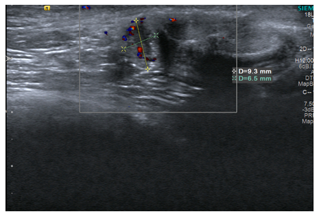
The patient underwent laparotomy and evaluated intraoperatively, and it was decided to continue chemotherapy. During this period, the patient was referred to our clinic again upon his complaint about a mass on his penis. On physical examination, an approximately 1-cm firm, fixed mass with a smooth structure was detected; therefore, superficial soft-tissue ultrasonography was performed, which revealed a solitary lesion of 9 x 7 x 6 mm (Figure 1). Since metastasis could not be excluded, penile magnetic resonance imaging (MRI) was planned for the patient. The MRI images showed an 8 x 7 x 5.5 lesion suspected to be malignant located in the dorsal of the penis at the middle level in the right half, which showed predominantly peripheral-style enhancement in the post-contrast examination (Figure 2). The lesion mostly originated from the buck fascia and was also associated with the cavernous body in the form of two thin vascular lines. The patient was taken into surgery under general anesthesia. A 1-cm skin incision was made from the area where the lesion was palpable. The mass, which appeared to be invading the buck fascia and the cavernous body, was totally excised. The cavernous body and buck fascia were repaired and the skin was closed. There was no postoperative bleeding or complication. The pathology result was adenocarcinoma metastasis. The patient continued to receive chemotherapy. On the first post-operative month, the superficial soft-tissue ultrasound of the patient showed that the operation site was clean, and there was no new involvement of the penis.

Figure 1: Ultrasonography image of the penis shows a hypoechoic mass.

Figure 2: MRI image of the penis shows the metastatic lesion.
Discussion
It remains unknown why penile metastases are seen very rarely despite the penis having a rich blood supply and an intense circulatory relationship with its neighboring organs [1]. Penile metastasis is a common disease symptom and a sign of poor prognosis [16]. The primary source of metastases is the bladder in 35% of cases, prostate in 30%, rectosigmoid in 16%, and kidneys in 6.5% [17]. Generally, 1/3 of the patients are diagnosed simultaneously with the primary tumor, while the remaining penile metastases are seen on average 18 months after the diagnosis of the primary tumor [18]. It has also been reported that there are late penile metastases seen many years after the primary tumor has been treated [19]. To the best of our knowledge, our patient is the fifth case with penile metastasis secondary to pancreatic cancer reported in the literature. The first case, reported in 1971 by Weitzner [6], developed due to pancreatic squamous cell carcinoma with multiple organ metastases. In 1989, Hashimoto et al. [13] presented a case of pancreatic head cancer with penile, prostate, and liver metastases. In 1997, Ahn et al. [14] reported the third case, in which multiple metastases were seen in pancreatic adenocarcinoma, and the patient, who refused radiotherapy for penile metastasis, died within a short period. The fourth case, reported by Virdis [15] et al. in 2019, belonged to a patient diagnosed with pancreatic cancer presenting with dorsal vein thrombosis and penile metastasis.
In conclusion, metastasis is very common in pancreatic adenocarcinoma; however, penile involvement is so rare that there is no standard treatment approach. Treatment options include local excision, penectomy, radiotherapy, and chemotherapy; however, there is an insufficient number of studies on the efficacy of radiotherapy and chemotherapy in this patient group. Since metastatic penile tumors are usually associated with advanced disease, the survival time after diagnosis is very short, with 80% of cases resulting in mortality within six months [20].
References
- Cherian J, Rajan S, Thwaini A, et al. Secondary penile tumours revisited. Int Semin Surg Oncol 2006; 3: 33.
- Song L, Wang Y, Weng G. Metastasis in penile curpus cavernosum from esophageal squamous carcinoma after curative resection: a case report. BMC Cancer, 2019; 19: 162.
- Chaux A, Amin M, Cubilla AL, et al. Metastatic tumors to the penis: a report of 17 cases and review of the literature. Int J Surg Pathol, 2011; 19: 597–606.
- ABESHOUSEBS, ABESHOUSE Metastatic tumors of the penis: a review of the literature and a report of two cases. J Urol, 1961; 86: 99-112.
- McCREALE, TOBIAS GL. Metastatic disease of the penis. J Urol, 1958; 80(6): 489-500.
- Weitzner Secondary carcinoma in the penis. Report of three cases and literature review. Am Surg, 1971; 37(9): 563-567.
- HayesWT, Young JM. Metastatic carcinoma of the penis. J Chronic Dis, 1967; 20(11): 891-895.
- MukamelE, Farrer J, Smith RB, deKernion JB. Metastatic carcinoma to penis: when is total penectomy indicated? Urology, 1987; 29(1): 15-18.
- Robey EL, Schellhammer Four cases of metastases to the penis and a review of the literature. J Urol,1984; 132(5): 992-994.
- Fischer MA, Patrick A. Secondary penile carcinoma from squamous cell carcinoma of the lung. Can J Urol, 1999; 6(6): 899-900.
- HillJT, Khalid Penile denervation. Br J Urol, 1988; 61(2): 167.
- Ryan DP, Hong TS and Bardeesy N. Pancreatic Adenocarcinoma. N Engl J Med, 2014; 371: 1039–1049.
- Hashimoto H, Saga Y, Watabe Y, et al. Case report: secondary penile carcinoma. Urol Int, 1989; 44: 56–57.
- Ahn TY, Choi EH, Kim KS. Secondary penile carcinoma originated from pancreas. J Kor Med Sci 1997; 12: 67.
- Virdis M, Bonifacio C, Brambilla T, Capretti G, De Nittis P, Uccelli F, et al. Thrombosis of the dorsal vein of the penis as first clinical presentation of pancreatic cancer metastatic to the penis. Tumori Journal, 030089161984927, 2019.
- Osther PJ, Lontoft E. Metastasis to the penis: Case reports and review of the literature. Int Urol Nephrol, 23: 161-167; 1991.
- Hızl F, Berkmen F: Penile metastasis from other malignancies. A study of ten cases and review of the literature. Urol Int, 2006; 76: 118-121.
- Pomara G, Pastina I, Simone M, et al: Penile metastasis from primary transitional cell carcinoma of the renal pelvis: First manifestation of systemic spread. BMC Cancer. 2004; 4: 90.
- Berger AP, Rogatsch H, Hoeltl L, et al: Late penile metastasis from primary bladder carcinoma. Urology, 2003; 62: 145.
- Demuren OA, Koriech O. Isolated penile metastasis from bladder carcinoma. Eur Radiol, 1999; 9: 1596-1598.

