Aesthetic Emergency in Fixed Prosthesis: Ceramo-Metallic Crowns’ Fracture Treatment
Didia EL1,*, Thioune N2, Ghoul S3 and Djeredou KB4
1Associate Professor in fixed prosthesis, International University of Rabat Morocco
2Master Assistant in fixed prosthesis, Cheick Anta Diop University Dakar Senegal
3University Professor, International University of Rabat Morocco
4Full Professor, Félix Houphouet Boigny University Abidjan Ivory Coast
Received Date: 13/06/2022; Published Date: 23/06/2022
*Corresponding author: DIDIA Ekow Léon Eric, Associate Professor in fixed prosthesis, International University of Rabat Morocco, College of Health Sciences, International Faculty of Dental Medicine, BioMed Unit, Morocco
Abstract
Introduction: Fixed prosthesis emergencies are mainly characterized by fractures and loosening. When a ceramic fracture occurs on the anterior crowns, the esthetic concern motivates urgently the patient to consult and usually requires from the dentist an immediate treatment in the office to urgently repair it. The repair of fractured restorations, a routine performance in some European countries, remains an unrecognized act in daily practice of developing countries.
In this clinical case, the authors report their experience in the emergency management of a 45-year-old patient presenting a medium-range bridge with a ceramic fracture on the lateral incisor crown.
The therapeutic decision was an aesthetic and functional rehabilitation of the fractured crown by chairside repair using the Ivoclar ® "Ceramic Repair" kit.
Clinical Case: On clinical examination, the patient presented a cervical fracture of the cervical third of ceramic-metal crown (CMC) on the lateral incisor (12) with an exposure of the metal and transverse cracks of the ceramic on the remaining two thirds.
The therapeutic decision was an aesthetic and functional rehabilitation of the fractured crown by chairside repair using the Ivoclar ® "Ceramic Repair" kit.
Discussion and Conclusion: The urgent management of a ceramic-metal fracture by this method is a wise choice. It corrects the aesthetic damage suffered at a lower cost by avoiding the loosening of the bridge, the removal of which is difficult or even impossible without sacrificing it.
Keywords: Ceramic-Metal Crown; Dental Prosthesis Repair; Metal Ceramic Alloys
Introduction
According to a study conducted by Proaño and Özcan in 2021 and 2002, when metal ceramic crowns are made, fractures begin to appear after a decade at a rate of 3-4%. On the other hand, ceramic crowns seem more resistant to fracking, as the first cracks appear after 10 years at a rate of 5 to 10%. In addition, 65% of fractures occur in the anterior sector, and in 75% of cases on the buccal surfaces of the maxillary teeth [8,1].
In case of a ceramic fracture of a prosthetic restoration, several therapeutic solutions exist. The practitioner can choose a total reconstruction or the repairing of the specific prosthetic part that is fractured. The aesthetic impact on the patient leads the practitioner to urgently perform acts of repair. Advances and progress in adhesion materials aimed at extending the life of our restorations in line with minimum principles of dentistry. Therefore, the objective of this article is to describe a protocol used to repair fractured ceramics at the dental office, by reconstitution using the "Ceramic Repair" kit from Ivoclar ®.
Presentation of the Clinical Case
We received a male patient, nurse, with a cervical fracture of a ceramic crown and financially unable to do it again immediately. The dental materials that were used to conduct this study were a Ceramic Repair® box, a photopolymerization lamp, a box of diamond cutters composed of oblong cutters with black and red rings, finishing and diamond cutters (extra fine grains).
Clinical (Figure 1A) and radiological examination (Figure 1C and 1D) of the fractured crown were conducted. The fracture occurred 4 years after initial treatment. The initial treatment was conducted with another practitioner, so the nature of the ceramic was not évident and the patient was not
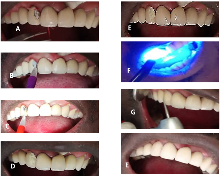
Possible premature contacts were examined throughout occlusion control using occlusion paper and eliminated (Figure 1B). The future shade of the final resin composite Tetric Evoceram® was determined among the proposed shades (Figure 1E).
The glaze of the veneering ceramic was removed from the margins of the repair using a fine-grit diamond rotatory bur with a shoulder edge (Verdent, Figure 1F) under water cooling. The bevel shape at the future bonding area was then created. The zirconia veneering and thereafter core surface were rinsed and conditionner monoband pus was applied on both metallic and ceramic surfaces for 60 seconds and air dried. Thereafter, the adhesive resin (Heliobond) was applied to the entire surface to be bonded and photopolymerized for 10 seconds using a photopolymerizing lamp (Figure 1G). The resin composite (Opaque®) was applied at 0.5mm and bonded to the conditioned surfaces and photopolymerized for 20 seconds (Figure 2A-G). The finall resin composite Tetric Evoceram® was applied and photopolymerized for 10 seconds. The finishing and polishing procedures were performed using bur for finishing (Verdent). Area was isolated using salivary rolls.
The satisfaction of the patient obtained, and the aesthetics regained, the control is carried out a month later, then three and six months later.

Figure 1: Initial situation of the patient and the Ceramic Repair material.
A: The endobuccal view of the fracture (The inspection of teeth
B: recording the laterality movements of occlusion
C et D : radiography of inlay core of the 22 and endodontic restoration of the 24
E: the kit Ceramic repair
F: dental diamonds burs for restoration
G : lamp of polymerisation

Figure 2 : Treament and final result.
A: Preparing the area to be repaired
B: Application of the conditioner Application of the binding agent (Heliobond®):
C: Application of the liaison opaque (IPS Empress Direct Opaque®)
D and E: Repair of the restoration by the TETRIC EVOCERAM® application
F: Photopolamerisation
G and E: polish and final result
Discussion
When considering the need to repair a chipping or core fracture directly, the size and location of the failure should be considered carefully, especially in multiunit restorations. Moreover, the prosthesis should be evaluated clinically and radiographically, as such complications might cause aesthetic problems and discomfort due to sharp edges.
The development of techniques for making a fixed prosthesis now makes it possible today to obtain prostheses usually considered as "definitive", because they are very precise from an aesthetic and functional point of view. However, despite respecting the different stages of development, no practitioner can claim to be immune from prosthetic failure [6]. This is valid regardless of the practitioner’s experience, rigor or skill. In addition, several studies have shown that all materials undergo a more or less severe attack. Saliva causes the alkali ions to disintegrate in the ceramic, resulting in the aggravation of the surface’s defects, and leading to cracks and fractures [7].
The advantage of this type of repair is its simplicity of implementation compared to the protocol of loosening and / or dismantling a bridge. It is rapid, performed in a single clinical session, reversible and does not require a laboratory phase. However, we have aesthetic and mechanical limitations. If the esthetic occlusal space or the free edge is insufficient to place a new restoration, the ideal is to repeat the design of the prosthetic part. Mechanical limits can be encountered and often related to shear forces and the relative weak seating of the anterior composite. This leads to the fracture of the ceramic at this level, hence the need to completely repair the prosthetic part. However, it should not be ruled out that repairing the ceramic does not always eliminate the etiology of the fracture because in fact, fractures can result, for example, from an undersized infrastructure, underlying the interest of a careful and well-conducted clinical examination to have a relatively long-lasting repair.
Conclusion
Ceramic fractures in restorations can occur accidentally following trauma (without warning signs). Their previous location responds to an aesthetic emergency for patients and practioners. The speed, the satisfaction of the patient, the possibility of performing this procedure for any dentist with a minimum of investment and immediate management of the aesthetic emergency seems to be justified for the adoption of this treatment in daily practice. Chairside crown repairs are lead dental surgeons to acquire these ceramic repair kits in their daily practice.
Disclosure: The authors have no financial interest in any of the companies or products mentioned in this article.
Conflicts of interest: the authors declare that they have no conflicts of interest concerning this article.
References
- Özcan M, Niedermeier W. Clinical study on the reason for and location of failures of metal- ceramic restorations and survival of repairs. Int J Prosthodont, 2002; 15: 299-302.
- Ugolini A, Parodi GB, Casali C, Silvestrini-Biavati A, Giacinti F. Work-related traumatic dental injuries: Prevalence, characteristics and risk factors. Dent Traumatol.. 2018; 34(1): 36-40.
- Sailer I, Makarov NA, Thoma DS, Zwahlen M, Pjetursson BE. All-ceramic or metal- ceramic tooth-supported fixed dental prostheses (FDPs)? A systematic review of the survival and complication rates. Part I: Single crowns (Scs). Dental Materials, 2015; 31(6): 603-623.
- Ohno H. New mechanical retention method for resin and gold alloy bonding. Dent Mater, 2004; 20(4): 330-337.
- Berthault GN, Durand AL, Lasfargues JJ, Decup F. Les nouveaux composites : évaluation et intérêts cliniques pour les restaurations en technique directe. Rev Odontostomatol, 2008; 37(3): 177-197.
- Anusavice K.J. Standardizing failure, success, and survival decisions in clinical studies of ceramic and metal-ceramic fixes dental prosthesis. Dental Materials, 2012; 28(1): 102-111.
- Archien C. Vita Enamic. Inf Dent, 2015; 97(29): 26.
- Lénine Proaño, Rebeca K Silva, Ariane Cc Cruz. A simple technique to repair the chipping of feldspar porcelain in a screw implant prosthesis: a clinical technique J Contemp Dent Pract, 2021; 22(1): 101-104.

