Synchronous Cardiac Myxoma and Lung Cancer in a Patient with Previous Prostate Cancer: A Rare Coincidence?
Houda Mokhlis*, Mohamed Malki, Reda Mounir, Meryem Boumaaz, Achraf Zaimi, Sara Ahchouch, Nadia Loudiyi, Hajar El Agouri, Najat Mouine, Ilyasse Asfalou and Aatif Benyass
Department of Cardiology, Mohammed V Military Hospital, Mohammed V University, Rabat, Morocco
Department of Cardiovascular Surgery, Mohammed V Military Hospital, Mohammed V University, Rabat, Morocco
Department of pathology, Mohammed V Military Hospital, Mohammed V University, Rabat, Morocco
Received Date: 17/05/2022; Published Date: 27/05/2022
*Corresponding author: Dr Houda Mokhlis, Department of cardiology, Mohammed V Military University Hospital, Rabat, Morocco
Abstract
Cardiac myxomas are the most frequent forms of primary cardiac neoplasms, it is a histologically benign tumor but it can block blood flow and cause other serious complications. Our case illustrates the concomitant occurrence of a left atrial myxoma that partially obstructed the mitral valve, and a primary lung cancer in a patient who underwent combined hormonal-radiation therapy for prostate cancer.
Keywords: Synchronous tumors; Cardiac Myxomas; Lung Cancer; Case Report
Introduction
Cardiac myxomas are the most frequent forms of primary cardiac neoplasms, with a percentage of 27% of all cardiac tumors [1]. Three quarters of myxomas form in the left atrium, It is a histologically benign tumor but can block blood flow and cause other serious complications.
On the other hand, Lung cancer is one of the most common and serious types of cancer. It is the leading cause of cancer-related death worldwide; accounting for approximately 20% of all cancer deaths [2] The association of two tumors of different origin in the same patient is becoming more and more frequent in the current clinical practice.
ere is presented the case of a patient who had a large left atrial myxoma that partially obstructed the mitral valve, as well as an unrelated, coexistent bronchial carcinoma and a history of prostate cancer.
Case Report
We report the case of a 57 years old male patient, a former smoker with a history of prostate adenocarcinoma treated in 2016 with full remission.
Four years later following the onset of a dry cough as well as a left basithoracic pain, the patient was diagnosed with an infiltrating bronchial squamous cell carcinoma and thus benefited from radio-chemotherapy sessions.
Months after initiating the therapy a chest CT scan was in favor of a good therapeutic response with a decrease in the size of the tumor, however it also showed a defect of opacification of the left atrium; a thrombus was suspected and the patient was urgently referred to cardiology consultation.
Clinical examination and EKG were unremarkable, Emergency TTE revealed a rounded heterogenous large mass with a smooth surface measuring (3.6 x 4cm) in the left atrium attached to the interatrial septum hindering ventricular filling with 7 mmHg as a mean transmitral gradient evoking a myxoma (Figure 1).
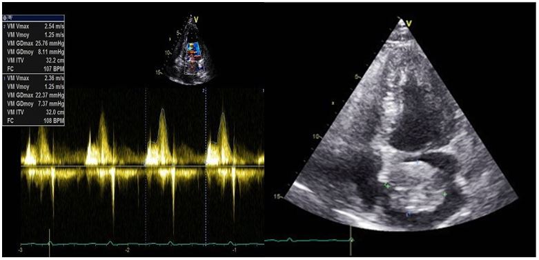
Figure 1: TTE images showing the mass.
However, TTE alone wasn’t enough to distinguish a benign primary from a metastatic cardiac tumor since it can’t give us a definite tissue histology; hence an MRI was performed showing a highly mobile masse, occasionally prolapsing through the mitral valve on SSFP sequences. On T1-w it was isointense and had higher signal intensity on T2-w. Late Gadolinuim enhancement sequences showed a heterogenous appearance (Figure 2).
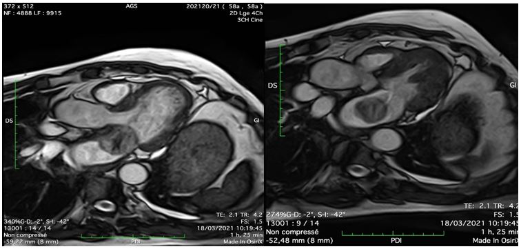
Figure 2: MRI images of the mass.
Decision-making, Therapeutic Intervention
Once a myxoma has been diagnosed, urgent surgical removal should be considered due to the risk of embolization, cardiovascular complications and sudden death; despite the poor prognosis of bronchial cancer our patient underwent urgent surgical excision (Figure 3) after consulting with the oncology team.
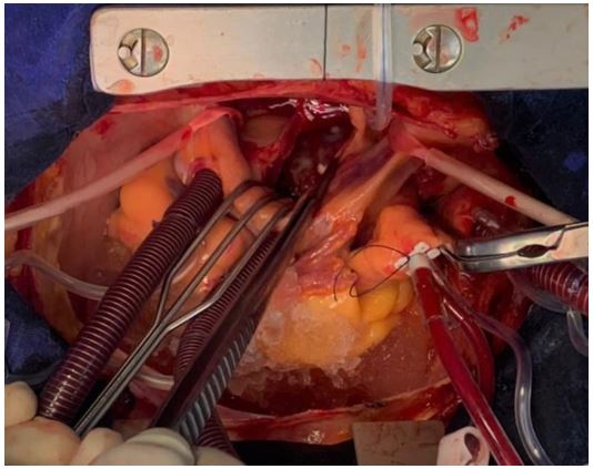
Figure 3: Surgical exeresis on the tumor mass.
Follow-up and outcomes
The surgical follow-up was simple; postoperative echocardiography confirmed complete excision of the mass and the anatomopathological exam confirmed the diagnosis of myxoma.
The excised gross specimen showed a 50×40×20 mm lobulated mass weighing about 38 g (Figure 4). The histological examination of the specimen revealed a lobulated mass covered by endothelium and composed of abundant loose myxoid stroma, containing abundant mucopolysaccharides. The myxoma is composed of scattered round, polygonal and stellate cells containing eosinophilic cytoplasm and uniform round or oval nuclei (Figure 5). These features are consistent with the diagnosis of atrial myxoma.
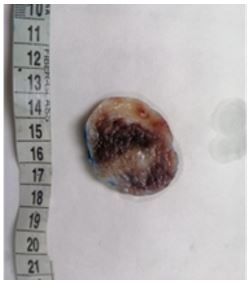
Figure 4: Gross specimen showing the lobulated mass excised from the left atrium measuring 50×40×20 mm. This cut section shows a soft myxoid surface with areas of haemorrhage.
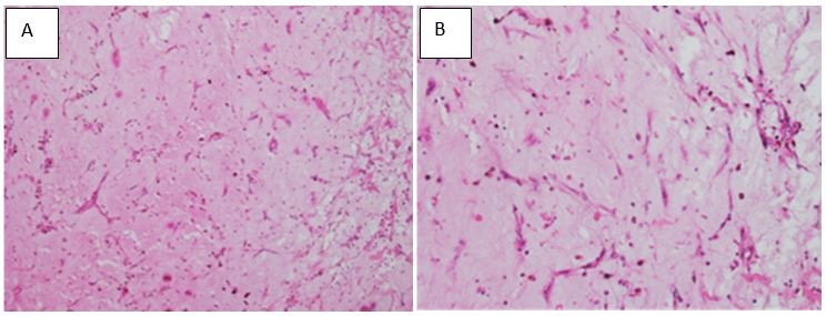
Figure 5: Histopathological examination showing abundant loose myxoid stroma with scattered round, polygonal or stellate cells with unirform oval nuclei. (Hematoxylin and eosin stain; A: magnification ×100, B: magnification ×200).
Discussion
Differentiating benign from malignant tumors is important for management and surgical planning. Cardiac myxomas are the most common benign tumor of the heart, usually arising in the left atrium.
The first-line examination, usually sufficient for diagnosis, is echocardiography. It can assess the presence of a hyperechoic mass, usually attached to the interatrial septum, with a heterogeneous texture in large lesions.
In doubt MRI is a very useful tool; myxomas demonstrate an intermediate but variable signal intensity on spin-echo images similar to that of the myocardium. they appear isointense on T1-weighted images and have higher signal intensity on T2-weighted images owing to the high extracellular water content. Regions of acute hemorrhage within myxomas appear hypointense on both T1- and T2-weighted images, and older regions appear hyperintense as the hemoglobin becomes oxidized to methemoglobin. Cine imaging is very useful in the work-up of myxomas as they are highly mobile, occasionally prolapsing through the mitral valve and causing obstruction. With SSFP cine techniques, myxomas appear hyperintense relative to the myocardium but hypointense relative to the blood pool. Contrast-enhanced sequences (first pass perfusion and LGE) are important in distinguishing myxomas from thrombus. Thrombi are avascular masses and therefore do not typically enhance on first pass and late perfusion sequences, however myxomas may contain cysts, regions of necrosis, fibrosis, hemorrhage, and calcification, which lead to a typically heterogenous appearance at contrast enhancement.
The description of multiple primary neoplasms dates from the late nineteenth century. Warren and Gates established the clinicopathological criteria for its diagnosis [3].
There is no established association between cardiac myxomas and lung neoplasms. Etiological factors for multiple primary cancers may include host and lifestyle-related factors, environmental and genetic factors and treatment related factors, in fact Multiple primary cancers can reflect the influence of previous cancer therapy, including chemotherapy and radiation therapy; genetic testing should be considered if an individual has two or more primary cancers with distinct histological features.
The association of a cardiac myxoma with a lung cancer is rare, with very few bibliography reports so far [4] ; however The medical literature describes cases of concurrence of cardiac myxoma and other malignancies; Kenan Iltumur [5] reported a casual echocardiographic finding of a left atrial myxoma that obstructed the mitral valve outflow tract, and an unrelated, concomitant cutaneous squamous cell carcinoma in the sacral area.
In 2018; Ernesto Chaljub Bravo [6] presented a case with a rare association of cardiac myxoma and hypernephroma, previously treated. Fibrolamellar hepatocellular carcinoma and cardiac myxoma are a rare presentation of synchronous tumors, there are at least two cases described of hepatocarcinoma synchronous with atrial myxoma [7-8].
Our case illustrates the concomitant occurrence of a left atrial myxoma and a primary lung cancer in a patient who underwent combined hormonal-radiation therapy for prostate cancer, to our knowledge, there are no previously published papers that describe this association.
Conclusion
Synchronous tumors are rare and the coexistence of malignant and benign tumors is even more so .Our case suggest that primary cardiac tumors which is a rare entity and other tumors can coexist however the relationship between the concomitant occurrence of a left atrial myxoma and cancer is unclear and this case may represent a pure coincidence thus further research is needed.
Conflicts of Interest: Authors do not declare any conflict of interest.
References
- Reynen K. Cardiac myxomas. N Engl J Med, 1995; 333: 1610–1617.
- Siegel RL, Miller KD, Jemal A. Cancer Statistics, 2018. CA: A Cancer Journal for Clinicians, 2018; 68: 7.
- Warren S, Gates O. Multiple primary malignant tumors. A survey of the literature and statistical study. Am J Cancer, 1932; 16: 1358-1414.
- Rahman MM, Ranjan R, Khan OS. Aftabuddin, Hoque MRS Left Atrial Myxoma in a Late Case of Lung Carcinoma (Mymensingh Med J, 2016: 25(2): 370-373.
- Kenan Iltumur, Tolga Demir, Zuhal Ariturk, Nizamettin Toprak, Oztekin Oto. Simultaneous occurrence of a large asymptomatic prolapsing left atrial myxoma with a cutaneous squamous cell carcinoma Heart Surg Forum, 2015; 18(1): E25-27.
- Ernesto Chaljub Bravo, Gustavo J, Bermúdez Yera, Alay Viñales Torres, Alain Allende González, Lisbetty López González, et al. Rare coincidence of two tumors: cardiac myxoma and hypernephroma.corsalud, 2018.
- Yessica M González-Cantú, Cristina Rodriguez-Padilla, Martha Lilia Tena-Suck, Alberto García de la Fuente, Rosa María Mejía-Bañuelos, Raymundo Díaz Mendoza, et al. Synchronous Fibrolamellar Hepatocellular Carcinoma and Auricular Myxoma Hindawi Publishing Corporation Case Reports in Pathology Volume 2015, Article ID 241708.
- Ochoa ML, Gordillo AGC, erez MMP, “Carcinomahepatocelular sincronico con mixoma auricular. Informe de uncaso,” Investigaci´on Cl´ınica, 2011; 52(2).

