Actinic Reticuloid: Case Report in a Moroccan Farmer
Mezni Line, Elhadadi Farah, Meziane Mariam, Ismaili Nadia, Benzekri Laila and Senouci K
Mohammed V University of Rabat , Ibn Sina University Hospital, Dermatology department
Received Date: 30/04/2022; Published Date: 20/05/2022
*Corresponding author: Mezni Line, MD, Department of Dermatology, Ibn Sina University Hospital, Av Mfadel Cherkaoui Souissi, 10100 Rabat Morocco
Abstract
Skin conditions arising on sun-exposed areas and worsening after exposure to the entire spectrum of solar radiation are getting more common and they represent diagnostic and therapeutic challenges. It was proven that ultraviolet radiation may have an impact on multiple molecular processes that damage the skin, inducing different skin changes and diseases. Thus, we report a case of Actinic Reticuloid (AR) as a rare dermatosis, encountering an increase in its prevalence.
Keywords: Actinic reticuloid; Chronic actinic dermatitis; Photosensitivity dermatitis
Introduction
Actinic Reticuloid (AR) is chronic photodermatitis caused by high photosensitivity to UVB, UVA and visible light beyond 400 nm. Lesions can vary from mild eczematous cases to AR and the most severe cases may mimic cutaneous T-cell lymphoma. The etiology and pathogenesis of the disease have not been fully established and the diagnosis is based on clinical, histopathologic, and photobiologic features [1].
Case Report
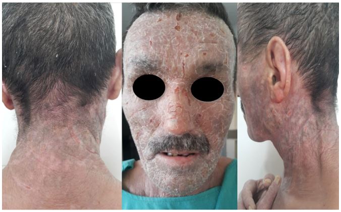
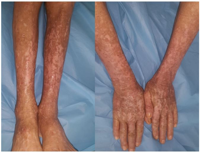
A 50-year-old Moroccan farmer with a 10 years history of a pruritic eruption that first appeared on the scalp, neck, upper back, forearms, and legs. He reported an exacerbation of the skin lesions while using medicinal plants and during the spring-summer period. No history of atopic diseases, drugs allergies, or daily use of colognes. On physical examination, we observed in sun-exposed skin: xerosis, erythematous scaly, indurated, achromic and hyperpigmented plaques. Excoriations, lichenification (Figure 1,2), an accentuation of the creases and a skin peeling on the face (Figure 1). Generalized lymphadenopathy was found on palpation and no abnormalities on blood tests. The diagnosis of actinic reticuloid was made based on the anamnesis, the clinical manifestations, the pathology and immunohistochemistry analysis of the skin and lymph node specimens. The patient was treated by hydroxychloroquine 200 mg, 1 tablet per day for three months, an emollient, a sunscreen SPF 50, and advised to wear sun-protective clothes. Unfortunately, he is lost to follow up.

Figure 1: Excoriation, skin peeling, dyschromia, hyperpigmented lichenified patches with an abrupt cutoff at the neckline.

Figure 2: Hyperpigmented and achromic patches with an abrupt cut-off at sun-shielded sites on legs and hands.
Discussion
Actinic reticuloid known as chronic actinic dermatitis, persistent light reaction, photosensitivity dermatitis, photosensitive eczema, photosensitivity and actinic reticuloid (PD/AR) syndrome is a skin condition that affects mostly elderly men, patients with a history of atopic dermatitis or contact allergies and younger patients with HIV infection. It frequently occurs in temperate climates and develops in Fitzpatrick skin type V and VI but all phototypes may be affected. It is hypothesized that the immune system overreacts to endogenous cutaneous antigens that are activated by solar radiation causing a similar reaction to allergic contact dermatitis. It has also been proven that allergic and/or photoallergic contact dermatitis commonly coexist with AR and often precedes the onset of photosensitivity (1). Therefore, the majority of AR patients have allergies to some substances which come into contact with their skin, particularly sesquiterpene lactones from compositae species, colophony, paraphenylendiamine, rubbers compounds, pesticides and sunscreens containing benzophenones [1,2].
Clinical manifestations display an eczematous eruption on sun-exposed areas and some features are highly indicative of the dermatosis such as hyperpigmented indurated plaques with an abrupt cutoff at sun-protected skin, and lichenification due to severe pruritus. In fact, lesions may extend to covered sites and some patients present a generalized erythroderma. Facial involvement in its severe form unveils leonine facies and the presence of localized and generalized lymphadenopathy may raise the possibility of cutaneous T cell lymphoma or other infiltrative processes [2].
Skin biopsy reveals non-specific patterns as epidermal spongiosis, psoriasiform hyperplasia, and superficial perivascular lymphohistiocytic infiltrate. Some atypical lymphocytes can be seen in the epidermis mimicking Pautrier's microabscesses. In this case, gene rearrangement analysis, in combination with immunohistochemistry are helpful in differentiating between AR and cutaneous T cell lymphoma. The papillary dermis is thick, with vertical collagen fibers and ectatic vessels. Some pathology hallmarks are important to consider, for instance, the presence of histiocyte-like cells with stellate cytoplasmic processes oriented toward the epidermal surface and in facial lesions the presence of a granulomatous component [3]. About two-thirds of patients with AR will have decreased minimal erythema doses to both (UVA) and (UVB) in photo-testing and patch- testing is considered when a contact dermatitis is suspected [4,5]. Hence, the diagnostic process is based on arguments in favor, excluding differential diagnosis as atopic dermatitis, mycosis fungoides, sezary's syndrome, tardive cutaneous porphyria and acute or subacute cutaneous lupus and by evaluating subsequently the ongoing therapy (1,2). Poor prognosis is related to severe sensitivity to UVB and/or the coexistence of a contact allergy. Treatment management is challenging, as it ranges from topical tacrolimus, low-dose PUVA, narrow-band UVB, B-carotene, hydroxychloroquine to immunosuppressive agents (eg cyclosporine, azathioprine, and systemic steroids (short time), mycophenolate mofetil) alone or in a combination regimen. Complete skin protection from (UV) light is crucial by using broad spectrum sunscreen that contains either Mexoryl SX or avobenzone as these filters provide the most effective UVA protection. Inorganic filters such as titanium dioxide or zinc oxide are efficacious but cosmetically unappealing [1,2,6].
Additional measures require patients to change their lifestyle such as seeking shade, avoiding direct sun exposure during the peak UV hours, the use of photoprotective clothing, wearing a wide-brimmed and avoiding provoking factors such as contact allergens [6].
Conclusion
Proper patient education is important to improve treatment outcomes as the disease has a chronic course causing a deterioration in quality of life. Nevertheless, drug therapy needs to be weighted and monitored as AR develops more likely in elderly patients who are at greater risk of suffering from adverse events as they are often poly medicated and have numerous comorbidities.
Funding source: none
Conflicts of interest: none disclosed
Consent for the publication of the patient’s photographs and medical information; Yes
References
- Lugović-Mihić L, Duvancić T, Situm M, Mihić J, Krolo I. Actinic reticuloid--photosensitivity or pseudolymphoma? A review. Coll Antropol. 2011; 35 Suppl 2: 325-329. PMID: 22220464.
- Hawk JLM. “Chronic actinic dermatitis”. Photodermatol Photoimmunol Photomed. 2004; 20: pp. 312-314.
- Sidiropoulos M, Deonizio J, Martinez-Escala ME, Gerami P, Guitart J. Chronic actinic dermatitis/actinic reticuloid: a clinicopathologic and immunohistochemical analysis of 37 cases. Am J Dermatopathol, 2014; 36(11): 875-881. doi: 10.1097/DAD.0000000000000076. PMID: 25238449.
- Yap LM, Foley P, Crouch R, Baker C. “Chronic actinic dermatitis: a retrospective analysis of 44 cases referred to an Australian photobiology clinic”. Australas J Dermatol, 2003; 44: pp. 256-262.
- Menage H du P, Ross JS, Norris PG, Hawk JLM. “Contact and photo contact sensitization in chronic actinic dermatitis: sesquiterpene lactone mix is an important allergen”. Br J Dermatol, 1995; 132: pp. 543-547.
- Paek SY, Lim HW. Chronic actinic dermatitis. Dermatol Clin, 2014; 32(3): 355-361, viii-ix. doi: 10.1016/j.det.2014.03.007. PMID: 24891057.

