Functional Rehabilitation of Mandibular Distal Extension Partial Edentulous Arch Combined with Maxillary Complete Edentulism
Neha Mukhopadhyay*, Sanjit Lal Das, Uttam Kumar Sen, Srijoni Mondal and Shrutokirti Banerjee
Department of Prosthodontics and Crown and Bridge, Haldia Institute of Dental Sciences and Research, India
Received Date: 18/04/2022; Published Date: 26/04/2022
*Corresponding author: Dr. Neha Mukhopadhyay, Post Graduate Trainee, Department of Prosthodontics and Crown and Bridge, Haldia Institute of Dental Sciences and Research, India
Abstract
Rehabilitation of a distal extension situation against maxillary edentulous ridge poses a challenging condition for the prosthodontist. Proper maintenance of function and aesthetics is difficult in these cases. Conventional fixed partial denture or implant-supported prosthesis is sometimes not feasible for distal extension cases due to unfavourable condition. In this case report, rehabilitation of a mandibular distal extension situation opposing a completely edentulous maxillary arch is described using removeable prosthesis using precision attachment.
Keywords: Distal extension; Attachment; Kennedy Class I; Preci-Vertex
Introduction
Successful prosthetic rehabilitation not only requires careful attention and meticulous treatment planning but also requires rehabilitating adequate aesthetics and function. Prosthodontic treatment options for replacement of missing dentition include Removeable Partial Denture (RPD), Fixed Partial Denture (FPD), and implant prosthesis. Rehabilitation of partially edentulous arch is a challenge, especially when it is a distal extension situation classified under Kennedy’s class I and class II situations [1]. Occlusal rehabilitation of distal extension case becomes even more difficult when it is opposing an edentulous arch.
For a distal extension situation, a fixed partial denture cannot be fabricated because of missing distal abutment. Implant-supported prosthesis can be planned, but it is sometimes not feasible due to unfavourable bone condition. In such situation an acrylic partial denture or a cast partial denture is largely preferred. Cast partial dentures are made retentive by the use of direct and indirect retainers and precision attachment components [2].
Attachments in prosthodontics could be extracoronal and intracoronal. Attachment-retained cast partial dentures facilitate both esthetic and functional replacement of missing teeth. Studies by various authors have shown a survival rate of 83.35% for 5 years, of 67.3% up to 15 years, and of 50% for upto 20 years [3,4].
This article describes a case report of a patient with mandibular bilateral distal extension Kennedy’s class I condition which is prosthetically restored by a cast partial denture retained using an extracoronal castable precision attachment (Preci-Vertex attachment system) against a maxillary single complete denture.
Case Report
A 59-year-old female patient reported to the Department of Prosthodontics and Crown & Bridge of Haldia Institute of Dental Sciences and Research with missing mandibular molars bilaterally (Figure 1: Showing Pre-operative Extraoral view). She gave a history of hypertension.
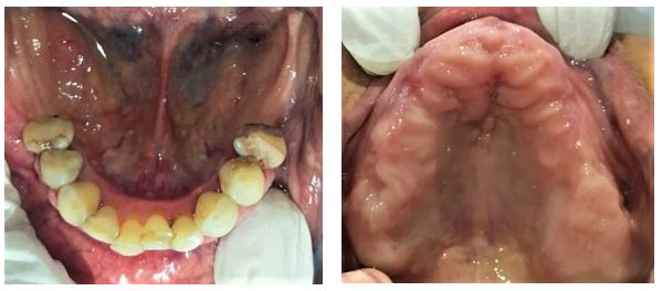
- On intraoral examination, it was noted that patient had missing mandibular first, second and third molars bilaterally (Kennedy’s Class 1) and she had endodontically treated (34,35,44,45). Also, she had completely edentulous maxillary arch (Figure 2: Showing Pre-operative Intraoral view).
- Diagnostic casts were made and mounted in tentative centric relation for diagnosis. Inter-ridge distance was measured. Clinical crown height was measured on 34,35,44,45.
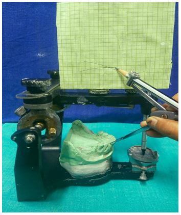
- Mandibular occlusal plane was determined using Brodrick’s Occlusal Plane Analyser. (Figure 3: Showing Brodrick’s Occlusal Plane Analyser).
- After complete clinical and radiographic examination, a prosthetic treatment plan was set up. A bilateral distal extension cast partial denture with extracoronal precision attachment on joint metal ceramic crowns wrt 34,35 and 44,45 was planned.
- An informed and written consent was obtained from the patient prior to initiation of treatment.
Clinical Procedure:
- Tooth preparation on 34,35, 44,45 was done to receive porcelain fused to metal (PFM) joint crowns attached to extracoronal attachment.
- Gingival retraction was done followed by impression making using putty and light body addition silicone material. (Densply Aquasil Addition Silicone) (Figure 4: Showing Tooth Preparation to Receive PFM Crowns wrt 34,35,44,45 followed By Cord Packing done Prior To Impression).
- Provisional restoration was looted.
Lab Procedure:
- Waxing up of abutments 34, 35, 44 and 45 was done and design of male component of attachment structure (PRECI-VERTEX) was waxed and then they were also cast along with the copings of the abutments (Figure 5: Showing laboratory wax up for joint metal coping with attachment).
Clinical Procedure:
- Metal try-in was done to check the overall fit of the copings and attachments. (Figure 6: Showing metal trial with male component of attachment).
- After metal trial was done, final porcelain fused to metal (PFM) joint crowns with extracoronal attachment on 34,35,44,45 was checked against the maxillary trial denture base for proper occlusion in centric relation. (Figure 7: Showing PFM crown with male component attached wrt 34,35,44,45)
Cast Partial Denture (CPD) Design and Fabrication:
- After proper planning and surveying, an appropriate cast partial denture framework was designed housing the female component of the attachment.
- The metal framework trial was done in the patient’s mouth for the accuracy of fit. (Figure 8: Showing cast partial denture framework trial done).
- Cast structure framework was checked up for stability and precision.
- Wax bite was taken after guiding the patient in centric relation position.
Wax-Up Trial:
- Tooth setting was done for mandibular cast partial denture.
- Waxing up of teeth was performed and teeth setting trial was done in patient’s mouth (Figure 9: Showing complete try in).
- The trial denture was sent for acrylisation and cast partial denture finished.
Prosthesis Insertion:
- Trial seating of the finished prosthesis was performed to check for fit, retention, occlusion.
- Cementation of joint crowns was done using Type 1 Glass Ionomer cement.
- Attachments were protected with a thin layer of petroleum jelly (Vaseline) in order to easily remove cast partial denture after joint PFM crowns with attachment have been seated.
- Female components were then inserted into the cast partial denture framework extraorally. (Figure 10: Showing PFM crown with male component luted, cast partial denture with female component).
- Complete seating of finished maxillary complete denture along with mandibular prosthesis with extracoronal distal extension precision attachment was done in the patient’s mouth. (Figure 11: showing maxillary and mandibular final prosthesis insertion - intra- oral)
- The patient was recalled after 24 hrs for post-insertion check-up.

Figure 1: Pre-operative- extra oral view.

Figure 2: Pre-operative- intra oral view.

Figure 3: Brodrick’s occlusal plane analyser.

Figure 4: Tooth preparation to receive pfm crowns wrt 34,35,44,45 followed by cord packing done prior to impression.

Figure 5: Laboratory wax up for joint metal coping with attachment.

Figure 6: Metal trial with male component of attachment.

Figure 7: PFM crown with male component attached wrt 34,35,44,45.

Figure 8: Cast partial denture framework trial done.

Figure 9: Complete try-in.

Figure 10: PFM crown with male component luted, cast partial denture with female component.

Figure 11: Maxillary and mandibular final prosthesis insertion - intra - oral.

Figure 12: Post-operative-intraoral frontal view (esthetics and occlusion restored).
Discussion
Precision attachment is a connector which consists of two or more parts. One part is connected to a tooth, root, or implant and the other part to the prosthesis providing a mechanical connection between the two. These attachments allowed prosthesis to combine the advantage of both fixed and removable restorations [5]. Dr. Herman Chayes who first reported the invention of attachment in the early 20th century [6].
GPT-9 defines precision attachment as a retainer consisting of a metal receptacle (matrix) and a closely fitting part (patrix); the matrix is usually contained within the normal or expanded contours of the crown on the abutment tooth/dental implant and the patrix is attached to a pontic or a removable partial denture.
Semiprecision attachment is defined as a laboratory fabricated rigid metallic patrix of a fixed or removable partial denture that fits into a matrix in a cast restoration, allowing some movement between the components; attachments with plastic components are often called semiprecision attachments even if prefabricated (not laboratory fabricated) [7].
Attachments give a removable prosthesis the exceptional feature of improved aesthetics, less postoperative adjustments, and better retention and improved comfort. (Figure 12: Showing esthetics and occlusion rehabilitation)
It is mostly indicated for long-span edentulous arches, distal extension bases, and nonparallel abutments [8]. There is a wide range of attachments available for different prosthodontic rehabilitation procedures from partial dentures to implant-supported prosthesis. By analysing study models and X-rays, the clinician can make several important points of determination, each of which will influence final attachment selection. Construction of such attachment require skill from dental technicians which cannot be acquired easily and needs training. The parts of the attachment are usually exposed to wear and tear and needed to be replaced over time [9].
PRECI VERTEX attachments system used in the case discussed in this article is extracoronal castable attachment positioned on the distal end of the crowns as an extension allowing a lot of vertical space. It is a very small attachment and requires minimal space. It provides optimal aesthetics with patient satisfaction. It is 4.5 mm height and may be reduced by 1 mm. The castable male component can be easily shaped together with the crowns during waxing-up stage avoiding complicated adaptation procedures like welding a metal attachment after crown casting. The male component design is cylindrical in shape with a flat head. The female component contains retentive nylon caps which are color-coded according to different retentive properties. Replaceable plastic female is available in three retention levels (white, yellow, red) and is incorporated directly into the framework. These nylon caps are replaceable and can be changed after wearing off.
Conclusion
Attachment retained removable prosthesis are a viable treatment modality for patients who cannot afford or are contraindicated for implant supported fixed prosthesis. However, lack of proper knowledge of the use of these attachments and inadequate training in this field leaves patients devoid of this treatment option [10].
References
- Mc Craken’s Removable partial denture prosthodontics 12th edition.
- Bakers JL, Goodkind RJ. Precision Attachment Removable Partial Dentures, Mosby, San Mateo, Calif, USA, 1981.
- Burns DR, Ward JE. “Review of attachments for removable partial denture design: 1. Classification and selection,” The International Journal of Prosthodontics, 1990; 3(1): pp. 98–102.
- Burns DR, Ward JE. “A review of attachments for removable partial denture design: part 2. Treatment planning and attachment selection,” The International Journal of Prosthodontics, vol. 3, no. 2, pp. 169–174, 1990.
- Preiskel HW. Precision Attachment in Prosthodontics, Quintessence Publishing, London, UK, 1995; 1-2.
- Preiskel HW. Precision Attachments in Prosthodontics: Overdentures and Telescopic Prostheses, Quintessence Publishing, Chicago, Ill, USA, 1985; 2.
- The Glossary of Prosthodontic Terms: Ninth Edition. J Prosthet Dent, 2017; 117(5S): e1-e105. doi: 10.1016/j.prosdent.2016.12.001. PMID: 28418832.
- Feinberg E. “Diagnosing and prescribing therapeutic attachment-retained partial dentures,” The New York State Dental Journal, 1982; 48(1): pp. 27–29.
- Makkar S, Chhabra A, Khare A, “Attachment retained Removable partial denture: a case report,” International Journal of Computing and Digital Systems, 2011; 2(2): pp. 39–43.
- Arora S, Anand S, Mittal S. Use of a semi-precision attachment to fabricate a removable partial denture. J Dent Specialities, 2017: 86-89.

