Calcifiing Odontogenic Cyst with Extensive Area of Bone Resorption Treated with Enucleation and Guided Bone Regeneration
Ariele Morgado Ribeiro¹,*, Erika Canabrava de Souza², Isabela Reis Mendes², Jôice Dias Côrrea²,¹,*
1Centro Universitário Presidente Tancredo de Almeida Neves, Uniptan, Brazil
2Pontífica Universidade Católica de Minas Gerais- PUC-Minas, Brazil
Received Date: 02/02/2022; Published Date: 14/02/2022
*Corresponding author: Jôice Dias Côrrea, Centro Universitário Presidente Tancredo de Almeida Neves, Uniptan, Pontífica Universidade Católica de Minas Gerais- PUC-Minas, Brazil
Abstract
The calcifying odontogenic cyst normally presents as a painless, slow-growing mass, involving most commonly maxilla, primarily the anterior segment (incisor/canine area). COCs represent a heterogeneous group of lesions that show a variety of clinicopathologic and behavioral features. Computerized tomography images revealed important characteristics that were not detected by panoramic radiography, as the expansion of the cortical bone. In this study, we report a case of intraosseous COC with extensive area of bone resorption treated with guided bone regeneration and perform an update regarding the characteristics of this cystic lesion and differential diagnosis, treatment, and prognosis.
Introduction
The Calcifying Odontogenic Cyst (COC) is a benign odontogenic cyst that occurs in the gnathic bones and was first delineated in 1962. This cyst is part of a spectrum of lesions characterized by odontogenic epithelium containing “ghost cells,” which may undergo calcification (Arruda et al, 2018) [1]. The Calcifying Odontogenic Cyst is a rare, its occurrence constitutes about 0.3–0.8% of all odontogenic cysts (Shear & Speight, 2007) [2] slow-growing lesion that has characteristics of both a cyst and a neoplasm, and therefore, in 2005, the World Health Organization (WHO) classified it as a Calcifying Odontogenic Tumor. Recently, this lesion was reclassified as a cystic lesion again by the WHO.
The same may be related to other tumors of odontogenic origin, such as ameloblastoma, odontoma and adenomatoid odontogenic tumors (NEVILLE et al, 2009) [3]. The documented literature confirms that that COC has a spectrum of variants, ranging from that of a developmental odontogenic cyst to benign and possibly a malignant odontogenic tumor (Shear & Speight, 2007) [2].
The aim of this article is to report a clinical case of a 60-year-old female patient diagnosed with Calcifying Odontogenic Tumor in the maxilla. This article also discusses the clinical and histopathological correlation of this condition and its impact on treatment/prognosis.
Case Report
A 60-year-old female patient presented in the private office for evaluation of a swelling on the alveolar ridge of the anterior mandible, as show in Figure 1, with the onset of symptoms approximately one year ago. During anamnesis, the patient reported to be hypertensive, controlled with the drug Captopril, 25mg. At the clinical examination, there was an increase in volume in the anterior region of the maxilla, hard to the touch and with a slight bluish color (Figure 1). The patient reported no painful symptomatology.
Computed tomography showed a large hypodense area between the roots of dental elements 12 and 13, causing distance between the roots and small hyperdense areas in the middle of the lesion as show in Figure 2.
First, radical endodontic treatment was performed on element 12. After 30 days with no sign of regression of the lesion, an excisional biopsy was performed, with an intrasulcular incision in elements 12, 13 and 14, with a distal relief incision and the mucoperiosteal tissue was detached.
In the transoperative period, in addition to surgical enucleation, an interposition of synthetic material for guided bone regeneration (Bio-OSS) were performed, considering the extension of the bone reabsorbed.
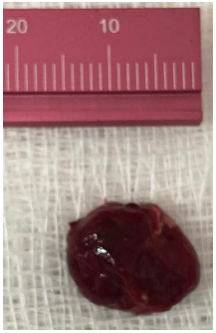
The entire lesion was removed by enucleation followed by curettage, where it was 15 mm in diameter, examination of the specimen revealed a firm and oval mass with cystic aspect, containing liquid in the interior (Figure 3).
The material was sent for histopathological examination in 10% formaldehyde solution. Microscopically, the hematoxylin and eosin (H and E) stained section showed a defined cystic lesion with a fibrous capsule and a lining of stratified epithelium outlining a matrix-producing stellate reticulate arrangement with calcifications.
The patient is under follow-up and after 2 months bone neoformation has been observed and there has been no clinical or radiographic evidence of recurrence (Figure 4).


Figure 2: Tomographic images of the lesion showing displacement of the roots of the right lateral incisor and canine, areas of bone resorption and expansion of the buccal and palatal cortical bone, in addition to points of calcification within the area of resorption.


Figure 4: Panoramic radiography revealing bone neoformation after 2 months of the surgical procedure.
Discussion
COCs represent a heterogeneous group of lesions that show a variety of clinicopathologic and behavioral features (Augustine et al, 2016) [4]. Originally described by Gorlin and colleagues in 1962 as a possible oral analogue to pilomatrixoma of skin, owing to the presence of ghost cell keratinization in both lesions. Nomenclature has been continuously changing, due to debate as to whether COC is a neoplasm or a developmental cyst. In 1992, WHO classified this lesion as an odontogenic tumor but continued to use the term calcifying odontogenic cyst. In 2005, WHO redesignated the lesion as calcifying cystic odontogenic tumor (CCOT) and in 2017, the term calcifying odontogenic cyst was reapplied to this lesion and it was reclassified as a benign odontogenic cyst (El-Naggar et al, 2017) [5].
Most cases are found in the incisor and canine areas (Buchner, 1991) [6]. Generally asymptomatic or a painless swelling. Radiographically, these lesions are usually an unilocular, well-defined radiolucency, although the lesion occasionally may appear multilocular. Radiopaque structures within the lesion, either irregular calcifications or tooth-like densities, may also be present in some cases. It can occur also in extraosseous regions, as in the gingiva, corresponding to 15 to 25% of all reported cases of COC (El-Naggar et al, 2017) [5].
COC represents 0.3% of odontogenic cysts, with no consistent gender or age predominance. May be associated with other odontogenic pathology, most commonly odontoma. Adjacent tooth root displacement, resorption or root divergence may occur (Arruda et al, 2018) [1].
Microscopically calcifying odontogenic cysts contain an ameloblastoma-like epithelial lining containing ghost cells that may calcify. Cyst wall consists of mature fibrous connective tissue containing scattered inflammatory cells (Arruda et al, 2018) [1]. Some studies associated COC with β catenin (CTNNB1) mutations (Yukimori et al, 2017) [7].
The treatment depends on the extension of the lesion. Most of the case could be removed by enucleation and curettage. Large cysts may be decompressed prior to surgical management in a 2-stage approach. For combined lesions, treat according to characteristics of more aggressive lesion. The prognosis is excellent, few recurrences (< 5% documented). However, although rare, malignant transformation has been reported (Motosugi et al, 2009; Zhu et al, 2012; Mokhtari et al, 2013) [3,8,9].
The differential diagnosis includes: Ameloblastoma, Dentinogenic ghost cell tumor / carcinoma, Ghost cell odontogenic carcinoma, Odontoma, Ameloblastic fibro-odontoma (Arruda et al, 2018) [1].
Conclusion
In summary, we report a case of an extensive anterior maxillary COC and demonstrate the importance of the correct diagnosis and that the treatment sometimes should not be limited to the removal of the lesion but also in the rehabilitation of areas of bone resorption that occur in lesions of great extension as reported here.
References
- Arruda JAA, Monteiro JLGC, Abreu LG, de Oliveira Silva LV, Schuch LF, de Noronha MS, et al. Calcifying odontogenic cyst, dentinogenic ghost cell tumor, and ghost cell odontogenic carcinoma: A systematic review. J Oral Pathol Med, 2018; 47(8): 721-730.
- Shear M, Speight P. Cysts of the oral and maxillofacial regions. 4th ed. Oxford, UK: Blackwell Publishing Ltd; 2007.
- Motosugi U, Ogawa I, Yoda T, Abe T, Sugasawa M, Murata S, et al. Ghost cell odontogenic carcinoma arising in calcifying odontogenic cyst. Ann Diagn Pathol, 2009; 13(6): 394-397.
- Urs AB, Augustine J, Singh H, Kureel K, Mohanty S, Gupta S. “Calcifying ghost cell odontogenic tumor (CGCOT) with predominance of clear cells: A case report with important diagnostic considerations,” Oral Surgery, Oral Medicine, Oral Pathology, Oral Radiology, and Endodontology, 2016; 121(2): pp. e32–e37.
- El-Naggar AK, Chan JKC, Grandis Takashi TJ, Slootweg PJ. Organization Classification of Head and Neck Tumours, World Health, Lyon, France, 2017.
- Buchner A. “The central (intraosseous) calcifying odontogenic cyst: an analysis of 215 cases,” Journal of Oral and Maxillofacial Surgery, 1991; 49(4): pp. 330–339.
- Yukimori A, Oikawa Y, Morita KI, Nguyen CTK, Harada H, Yamaguchi S, et al. Genetic basis of calcifying cystic odontogenic tumors. PLoS One, 2017; 12(6): e0180224.
- Zhu ZY, Chu ZG, Chen Y, Zhang WP, Lv D, Geng N, et al. Ghost cell odontogenic carcinoma arising from calcifying cystic odontogenic tumor: a case report. Korean J Pathol, 2012; 46(5): 478-482.
- Mokhtari S, Mohsenifar Z, Ghorbanpour M. Predictive factors of potential malignant transformation in recurrent calcifying cystic odontogenic tumor: review of the literature. Case Rep Pathol, 2013; 2013: 853095.

