Peri-Implant Fracture in an Operated Case of Left Sided Radius Ulna Shaft Fracture with Volar Plate - A Case Report and Review of Literature
Dr Neetin Mahajan, Jayesh Mhatre*, Dr. Pravin Sarkunde
Department of orthopedics, Grant govt medical college, India
Received Date: 03/01/2022; Published Date: 14/01/2022
DOI: 10.46998/IJCMCR.2022.17.000410
*Corresponding author: Jayesh Mhatre, Department of orthopedics, Grant govt medical college, Mumbai, India.
Abstract
We report a case of a 72-year-old male with a peri-implant fracture located just below to the plate and a fracture of the radius ulnar shaft occurred after a simple fall. The patient was surgically treated by plate and screws removal. The fracture was fixed using a 3.5 mm A peri-implant fracture near the plate of shaft of radius ulna represent a very rare injury. The main factor of this lesion is high energy trauma on the dcp for the radial and ulnar shaft fracture. Different factors such as osteoporosis, BMI and screw position could influence the fracture pattern.
Introduction
Forearm fractures are amongst the most common fractures in the adult population1, in this article, we describe a rare case of a peri-implant fracture due to a trauma near the volar plate of radius ulna shaft fracture which was successfully treated without long-term complication.
Case Presentation
A 72-year-old male attended to our emergency room after a fall on his left forearm. The patient was in good health with no known comorbidities, his medical history showed that 1 years previously the patient had undergone on an open reduction and internal fixation with plating for shaft of radius ulna fracture. The patient reported good clinical outcome without any local complications.
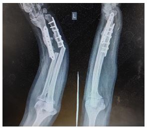
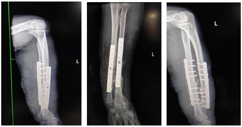
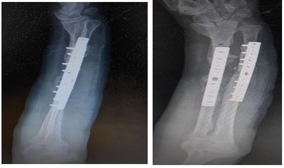
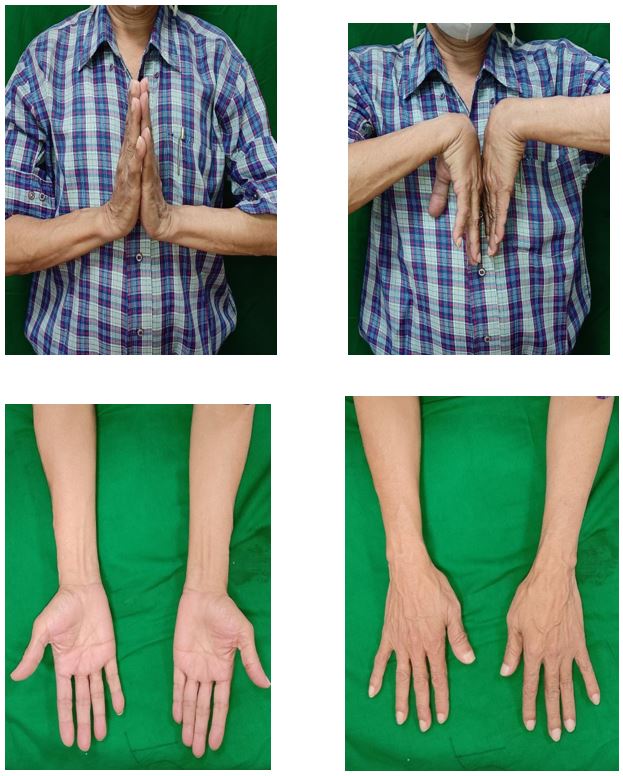
At the physical examination the forearm dorsal Angulation showed deformity. The patient was unable to actively move his forearm, with aggressive pain at passive movements. No vascular or neurological deficiencies were found. An X-ray examination revealed a peri-implant fracture of the radius and ulna shaft just below plate the plate was still affixed to shaft of radius and ulna (Figure 1). The following this, the patient was surgically treated. A modified henrys approach for radius shaft and subcutaneous approach for ulnar shaft was performed with a skin incision placed over the previous surgical scar. The plate and screws were removed, and the reduction was performed. Internal fixation was performed using a 3.5 mm dcp for both radius ulna shaft. Reduction maintenance and articular alignment were restored. The x-rays images after the procedure showed a good reduction of the fractures (Figure 2a). Postoperatively, the forearm was splinted for 10 days. After removing the stiches, the patient started rehabilitation to improve the range of motion in the forearm and wrist, using active and gentle active-assisted exercises. Clinical and radiological (Figure 3a) follow-up was scheduled at 1, 3 and 6 months. At the 30-day follow-up, the ROM was 25° of dorsiflexion, 30° of palmar flexion, 45° of pronation and supination.


Discussion
The peri-implant radius ulna shaft fractures are very rare, but probably the number of these fractures will increase more and more due to incremental use of plates for the treatment of fractures2. In the literature, only one similar case of fracture occurred proximally to the volar radius plate is reported. In this case, the radius fracture was associated with an ulnar shaft fracture, the patient was treated with the old implant removal and a new internal fixation with plate for the radius and a plate to the ulnar fracture3. in this case a simple fall was enough to create the fracture. The cause of this is probably attributable to the high degree of osteoporosis of the patient: the poor bone quality can compromise strength and eventually lead to bone fragility. In fact, osteoporotic bone is characterized by thinned cortex, decreased trabecular density, and decreased stiffness4,5. Further, it could be observed a resistance reduction at the level of the tip of the osteosynthesis implant that could act as a stress riser. Consequently, peri-implant fractures could occur as complications of osteoporotic fracture treatment
Analyzing the hardware construction, we observed that cortical screws were inserted in the proximal hole of the plate both cases. A recent biomechanical study shows that longitudinal shaft fracture and peri-implant fractures seemed relevant problem using locking screws in the proximal holes rather than cortical screws. For this reason, the cause of these atypical fractures must be sought in others mechanical problems. In literature is widely accepted that the midline of the bone is the best site for fixation, since screws have an optimal bi-cortical hold at this level. When a screw track is completely in cortical bone, more bone has been drilled through increasing the risk of fracture6. Analyzing the proximal screw position of the primary implant in our case. it is clear that the position of the screw was not perfectly placed in the midline of the bone, but it was in a “off-centre” position on the transverse plane. That screw position probably created a locus Minoris Resistencia of the radius shaft. Therefore, despite the simple fall, severe osteoporosis in addition to the high weight of the patient, increased the load near that weak point proximally to the plate creating the shaft fracture. Thus, with this type of fracture, it is necessary to investigate the presence of osteoporosis through blood tests and DEXA examination7 and set an adequate therapy. It will also be necessary to monitor the healing process of the fracture, periodically evaluating the disease parameters to follow the effectiveness of therapy
Conclusion
A peri-implant fracture near the volar plate of radius ulna shaft represents a very rare injury. The increase of the use of plates and screws could increase the incidence of this type of fracture. A trauma of high energy associated with poor bone quality can determine a new fracture around the plate.
References
- Wilcke MK, Hammarberg H, Adolphson PY. Epidemiology and changed surgical treatment methods for fractures of the distal radius: a registry analysis of 42,583 patients in Stockholm County, Sweden, 2004–2010. Acta Orthop, 2013; 84: 292–296.
- Court-Brown CM, Caesar B. Epidemiology of adult fractures: a review. Injury, 2006; 37:691–697.
- Jupiter JB, Marent-Huber M. LCP Study Group Operative management of distal radial fractures with 2.4- millimeter locking plates: a multicenter prospective case series. J. Bone Joint Surg, 2009; 91: 55–65.
- Arora R, Gabl M, Erhart S, Schmidle G, Dallapozza C, Lutz M. Aspects of current management of distal radius fractures in the elderly individuals. Geriatric orthopaedic surgery & rehabilitation, 2011; 2: 187–194.
- Thorninger R, Madsen ML, Wæver D, Borris LC, Rölfing JHD. Complications of volar locking plating of distal radius fractures in 576 patients with 3.2 years follow-up. Injury, 2017; 48: 1104–1109.
- Diaz-Garcia RJ, Oda T, Shauver MJ, Chung KC. A systematic review of outcomes and complications of treating unstable distal radius fractures in the elderly. The Journal of hand surgery, 2011; 36:824–835.
- Snoddy MC, A TJ, Hooe BS, Kay HF, Lee DH, Pappas ND. Incidence and reasons for hardware removal following operative fixation of distal radius fractures. J Hand Surg, 2015; 40: 505–507.

