Sacciform Aneurysm of Proximal Anterior Interventricular Artery Associated with a Tight Lesion of Mean Segment : Clinical Case Report and Literature Review
Larsen Clarck Moumpala Zingoula*, Asmaa Ameur, Chaimaa Rhemimet, Soumaré Ahamadou, Safae Malhouf, Fatine Tabti, Meryem EL Harrak, Nadia Fellat, Rokya Fellat
Department of Cardiology, National League for the Fight against Cardiovascular Diseases, Ibn Sina University Hospital, Rabat, Morocco
For our university: Mohammed V University of Rabat, Faculty of Medicine and Pharmacy
Received Date: 27/12/2021; Published Date: 13/01/2022
DOI: 10.46998/IJCMCR.2022.17.000409
*Corresponding author: Larsen Clarck Moumpala Zingoula, Department of Cardiology, National League for the Fight against Cardiovascular Diseases, Ibn Sina University Hospital, Mohammed V University of Rabat, Faculty of Medicine and Pharmacy, Rabat, Morocco.
Summary
Coronary aneurysms are rare vascular pathologies of various etiologies, the most common of which is atherosclerotic, most often associated with atheromatous lesions. Frequently asymptomatic, coronary aneurysm is either a fortuitous discovery on angiographic exploration for another cause. We report the clinical case of a patient in whom the diagnosis of coronary aneurysm was made accidentally during coronary angiography following the exploration of a coronary syndrome.
Keywords: Interventricular artery; Sacciform aneurysm; Coronary angiography
Introduction
Coronary Aneurysm (CA) is a well-described, rare condition, initially reported post-mortem, but increasingly reported since the era of cardiac catheterization. In most of the series reported, the incidence in patients screened for ischemic heart disease is between 1 and 2-5%. Associations of other localizations have been reported, such as aortic aneurysms with no obvious relationship. It is frequently associated with atherosclerotic coronary artery disease 1. Hence the interest we attach to this subject by reporting the clinical case of our patient in invasive cardiac exploration fortuitously objective an AC associated with a tight lesion of the artery average interventricular (VIA).
Clinical Case
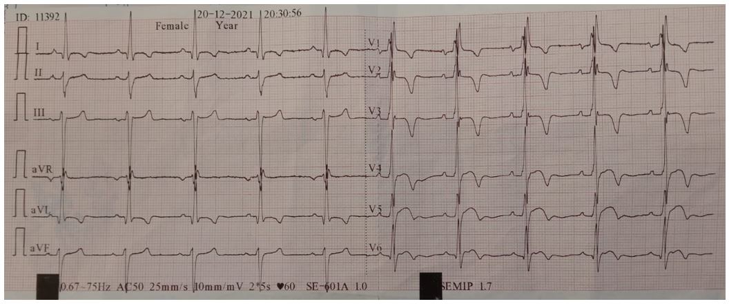
This is a 67-year-old patient, chronic smoking weaned for 3 years, who as part of her hypertension routine, evolving for 20 years under dual therapy, performs an ECG has a regular sinus rhythm with an elevation of ST apicolateral with hollow T waves in the extended anterior, diphasic T waves in the inferior and a left anterior hemiblock. Her admission laboratory workup was unremarkable, with troponin at 3 times normal and echocardiography revealed anteroseptal hypokinesia.
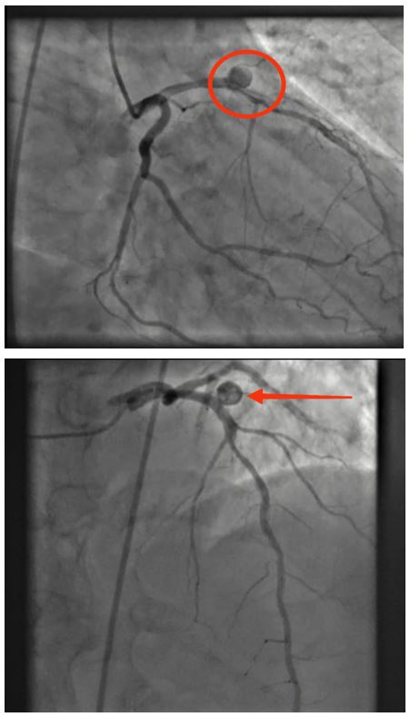
Coronary angiography revealed a sacciform aneurysm of the proximal Interventricular Artery (VIA) followed by tight stenosis of the middle VIA with a good downstream bed.


Discussion
Coronary aneurysm remains a less and less reported disease to this day despite the era of coronary angiography. Predominantly male, its overall incidence is 1 to 4% in patients whose coronary arteries are explored on coronary angiography [1].
We can distinguish sacciform aneurysms, spindle-shaped aneurysms and true aneurysms which involve the three arterial tunics.
Following the example of the anatomopathological study carried out by John E. Markis et al [2] on coronary aneurysms which revealed the following characteristics: diffuse hyalinization, deposition of lipids, destruction of the intima and of the media, focal calcification, etc. intima and media, focal calcification and fibrosis, cholesterol crystals, intramural hemorrhage and giant cell foreign body reaction, and giant cell foreign body reaction of the atherosclerotic process extending to the observing media extensive destruction of musculo-elastic elements resulting in marked attenuation of the vessel wall. In areas where the media is relatively intact, ectasia was absent.
It is therefore necessary to qualify, between aneurysmal dilation and poststenotic coronary ectasia is a well-known phenomenon. And that the coronary angiography of our patient reports a severe stricture of the average VIA which supports the atherosclerotic process in our patient whose cardiovascular risk factors are smoking, android obesity and hypertension. These last three remain independent factors of aneurysm progression [2].
Atherosclerosis remains the main etiology of coronary aneurysms, unlike false aneurysms whose causes are either congenital, or traumatic, syphilitic, bacterial or autoimmune in the context of Kawasaki-type vasculitis which is however a common cause in Asian countries [3].
Asymptomatic, coronary aneurysms are often diagnosed incidentally during a coronary examination in a patient explored for a coronary emergency, such is the case of our patient who in the course of an ischemic appearance found on his ECG was the subject of an invasive exploration [4].
As for diagnostic explorations, Coronary angiography remains the cornerstone providing more details in terms of the size, shape, location and number of the aneurysm. Also, the Coroscanner as well as the cardiac MRI which analyzes, in addition to the coronaries, the kinetics and the measurement of the right and left ventricular ejection fractions [4].
The complications are of a thromboemboligenic nature: there may be cases of myocardial infarction without significant lesions found on the coronary angiography except the aneurysm. This being the case, the intracoronary embolus of aneurysmal origin is the most probable hypothesis. This would justify the administration of long-term anticoagulant therapy (antivitamin K). Also, complications on rupture of a coronary artery aneurysm, which is extremely rare. Coronary aneurysm can rupture into the pulmonary artery, right ventricle, and coronary sinus and cause arteriovenous fistula with left-to-right shunt, hematoma, or intramyocardial mass. As for their rupture in the pericardial space would cause a pericardial tamponade [4].
The management for some authors is by endoprosthesis. However, Paul S. Swaye et al [4] did not show a difference in five-year survival, but a significantly higher rate of myocardial infarction in patients with aneurysm. As our patient, despite the proximal VIA aneurysm, has a tight lesion of the middle VIA that underwent stent revascularization.
Surgery is recommended to prevent complications. Ueyama K. et al [5] recommended surgery for patients with coronary aneurysms larger than 30 mm in diameter.
Conclusion
References
- Hartnell GG, Parnell BM, Pridie RB. Coronary artery ectasia; its prevalence and chirurgical significiance in 4 993 patients. Br Heart J, 1985; 54: 392-395.
- Markis JE, Joffe CD, Cohn PF, Feen DJ, Herman MV, Gorlin R. Clinical significance of coronary arterial ectasia. Am J Cardiol, 1976; 37: 217-221.
- Bernard F, Revel F, Monségu J, Duriez P, Langlade S, Ollivier JP. L’ectasie des vaisseaux coronaires: une coronaropathie à haut risque thrombo-embolique. Ann Cardiol Angeiol, 1998; 47(3): 160-164.
- Swaye PS, Fisher LD. Aneurysmal coronary artery disease. Circulation, 1983; 67(1): 134-138.
- Ueyama K, Tomita S, Takehara A, et al. A case of surgical treatment of cardiac tamponade caused by a ruptured coronary aneurysm accompanied by a coronary artery-pulmonary artery fistula. Kyobu Geka, 2001; 54(1): 70–75.

