Non Hodgin B Cell Gastric Lymphoma –A Case Report
Marsela Shani*, Griselda Pelo, Jerina Todhe
Prison Health Hospital, Mother Teresa University Hospital, Tirana, Albania.
Prison Health Hospital, Mother Teresa University Hospital Tirana, Albania.
Nr 1 Healthcare Center Tirana, Albania
Received Date: 15/11/2021; Published Date: 13/12/2021
*Corresponding author: Marsela Shani, Prison Health Hospital, Mother Teresa University Hospital, Tirana, Albania.
Lymphoma is a malignant tumor of lymphoid tissue. Lymphoid tissue is dispersed throughout the whole of the gastrointestinal tract; however, only in the upper aerodigestive tract and in the terminal ileum is it organized in structures similar to those present in other secondary lymphoid organs such as the spleen and the lymph nodes. In most of the cases the organization of the lymphoid tissue in functional structures is unnecessary because intestinal transit is rapid and the environment is not so favorable to microorganisms. In specific situations when there is presence of an inflammation, infection or a tumor, it is possible for persistent antigenic stimulus to favor the development of lymphoid tissue organized in such a way that it resembles mucosa-associated tissue present in other locations. In normal conditions, this lymphoid tissue can be observed in the cecal appendix. There have been described three types of cellular present in the lymphoid tissue of these organs. The first, made of true lymphoid aggregates of the mucous membrane with germinal centers, mantle zones, and a crown of peripheral lymphocytes called "marginal zone" lymphocytes. The second, labels the lymphoid tissue dispersed throughout the lamina propria, and the third includes the intraepithelial lymphocytes. There may be found different conditions that alter the normal structure of lymphoid tissue. Processes such as hyperplastic phenomena, lead to inflammatory processes that are followed by enlargement of normally present lymphoid tissue. There are also conditions which increase the T lymphocyte population. These include certain immunological processes and Helicobacter pylori infections in the stomach. Consequently, under certain conditions such as enduring stomach infection by Helicobacter pylori neoformation of an organized lymphoid system occurs and results in the formation of true lymphoid follicles Primary gastric lymphoma is a general term for a type of cancer that originates within the stomach.
Case Report
Patient F.P, male ,22 years old, had approximately one month with physical weakness, abdominal pain, intermittent temperature (38°C), vomiting. He was consulted by an abdominal surgeon and was hospitalized as suspected intestinal sub occlusion.
Life history: Regular smoker 20-40 cigarettes for 4-5 years.
Family history: Nothing important
Objective examination: CV system FC =82 bpm, TA 110/70mmHg, no pathological noises, normal ECG
Vital signs: Temperature 38.1°C, Sat O₂ 98%, smooth bronchovesecular respiration without pathological noises in both pulmonary areas, soft abdomen, without collateral vasal network, palpable liver and spleen under the costal arch, light muscular defense, without peritoneal reaction, colored skin and mucous membranes, with no subcutaneous icter.Genitourinary tract normal.Normal reflexes present with no pathological findings.
Blood count: RBC 4 320 000/mm³, Hbg 11.1 gr/dl,WBC 10.3x10⁹/L,(Gran 7.9x10⁹/L), MCv 80.4fL,PLT 429x10⁹/L
Biochemical values: Glucose 87mg/dl,Urea 24mg/dl,Creat 0.7 mg/dl,AST 45 U/L,ALT 23U/L, Amylase 45U/L, Total Bilirubine 0.3mg/dL,Total Proteine 7.6 g/dL,Na 134 mmol/L,K 3.7mmol/L,Cl 101 mmol/L. CRP 51.7 ,ERS 48mm/h,Stool antigen test positive for Helicobacter Pylori.
Abdominal ultrasound: Liver and bile ducts in normal. Pancreas with normal ecostructure, both kidneys with normal structure, urinary bladder emptied with normal ecostructure, without free fluid in the abdomen. Hyperperistaltic and intestinal distention are noticed.
CT abdomen: Trachea and main bronchi normal. Heart with normal dimensions without pericardial fluid. No mediastinal lymph nodes are observed, pulmonary fields with normal ventilation. Free phrenic-costal sinuses. Liver without obvious focal lesions. Cholecysts with thin walls without calculi inside. intrahepatic biliar ways are not enlarged. Empty stomach with thickening wall in gastroesophageal junction up to 28mm which contrasts markedly after injection of venous contrast. Locoregional and periaortical lymph nodes up to 8 mm. Densification of surrounding adipose tissue. Homogeneous spleen. Pancreas with normal dimensions, without dilated pancreatic duct.
Fibrogastroscopy: Esophagus with free passage. In the cardio-esophageal region is observed ulcerative lesion with irregular shape that extends more posteriorly by touching the lower esophageal sphincter (biopsy was taken). Z line with irregular shape.The rest of the stomach, normal ,without lesions. Duodenal bulb normal passage up to D2, without lesions.
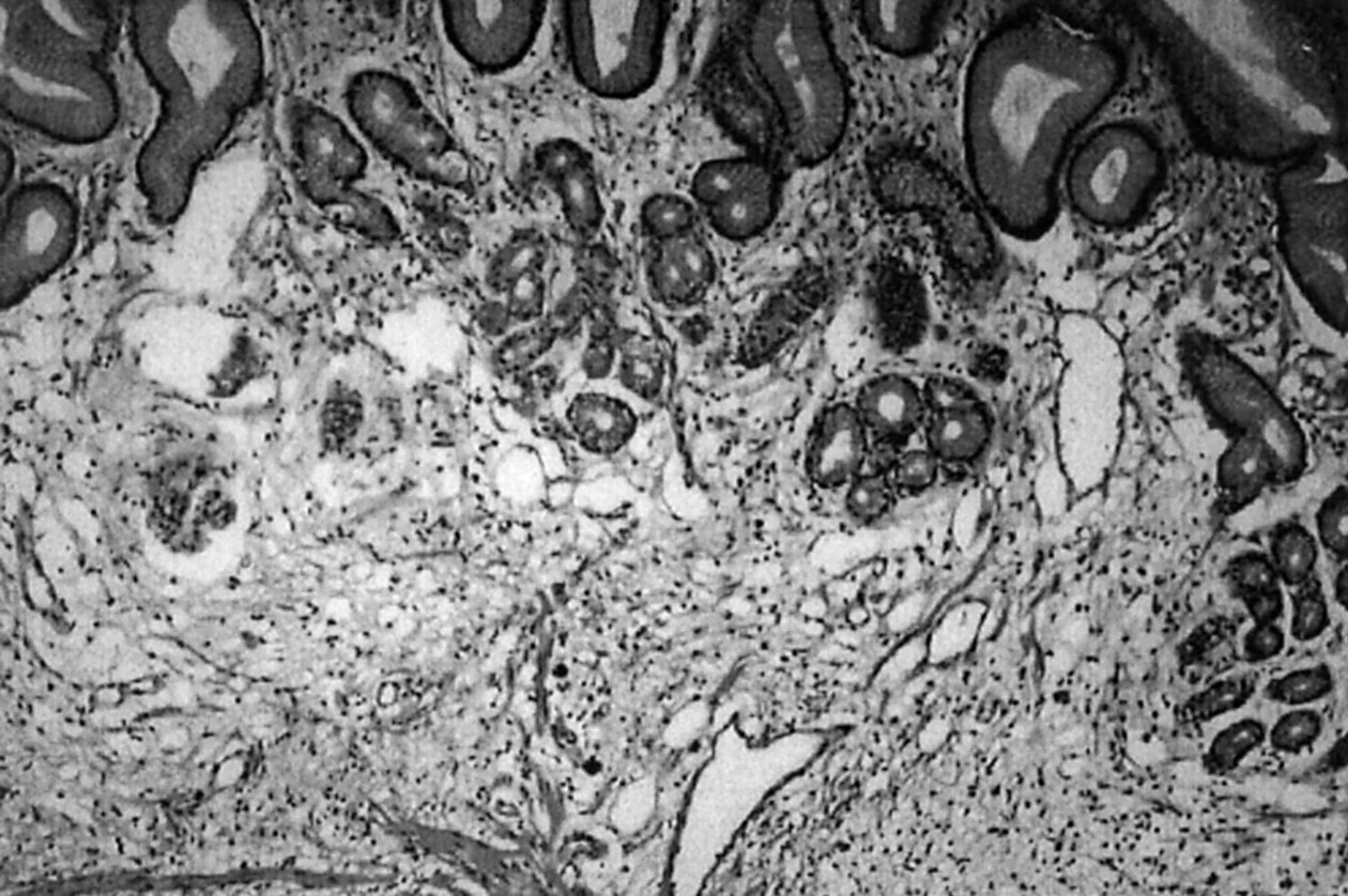
Biopsy of the material: In macroscopic examination, variegated particles are observed. All material was sampled. In the examined material, the gastric mucosa infiltrated by a malignant neoplastic lesion composed of basophilic cells that are organized in solid islands. To differentiate a lymphoma from malformed carcinoma, the sample was subjected to immunohistochemistry. Immunohistochemical examination resulted in PAN Keratin Neg (-), CD20 position (+) in atypical cells, CD45 position (+) in atypical cells. This profile supports the diagnosis of Non Hodgin lymphoma with B cells.

Therapy: The patient started therapy according to the R-CHOP scheme at 21-day intervals. The therapy-related compliance was good.
Primary Gastric Lymphoma (PGL) is the most common extranodal non-Hodgkin lymphoma and represents a wide spectrum of disease, ranging from indolent low-grade marginal zone lymphoma or mucosa-associated lymphoid tissue (MALT) lymphoma to aggressive diffuse large B-cell lymphoma. Chronic gastritis secondary to Helicobacter pylori (H pylori) infection has been considered a major predisposing factor for MALT lymphoma.
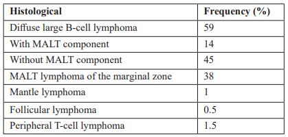
Distribution of the Main Histological Types (Defined According to the Criteria in the REAL Classification) in the German Multicenter Perspective Study for Gastrointestinal NHL. (Koch et al, 2005)

Clinical Presentation
The initial symptoms of PGL are usually nonspecific, mimicking gastritis, peptic ulcer disease, pancreatic disorders, or functional disorder of the stomach. On the physical examination usually, there is nothing specific in 55% to 60% of cases. These nonspecific findings affect the accurate and timely diagnosis of this pathology, which can be several years in certain cases. The most common symptoms reported include weight loss, nausea, vomiting, abdominal fullness, and indigestion. Weakness, night sweats, jaundice, fever, and dysphagia occur less frequently. Other uncommon symptoms are gastric obstruction and perforation, fever, hepatomegaly, splenomegaly, and lymphadenopathy. About 20% to 30% of patients with gastric DLBCL report gastrointestinal bleeding in the form of hematemesis or melena. In some cases, physical examination findings including epigastric tenderness, adenopathy, and palpable epigastric masses.
Pathogenesis of MALT Gastric Lymphoma
Association with chronic H pylori
Various studies have shown that about 75% of H pylori-positive gastric MALT lymphomas make a complete remission after eradication of this bacteria with antibiotic therapy, supporting the link between H pylori infection and the presence of MALT. When lymphoid cells are constantly stimulated by H pylori, they can cause MALT lymphoma. In addition to B cells, T cells and macrophages play an important role in MALT lymphogenesis. Over time, B cell clones that still depend on antigens for growth and survival, carrying unknown mutations, will appear. At this stage, the spread is monoclonal, but is not yet able to spread beyond the site of inflammation. Upon receipt of additional mutations, including chromosomal abnormalities, the tumor becomes antigen-independent and capable of systematic proliferation.In MALT gastric lymphomas negative H pylori, the theory that the infection leads to lymphomageneesis and the presence of lymphoma loses its validity. Today, there are many theories related to the idea that there are various mechanisms by which pathogenesis occurs in gastric MALT negative for H pylori, including the relationship between genetic changes (t (11; 18)) and other routes of activation.Primary gastric lymphoma is the most common form of extranodal localization in the NHL, representing about 30-40 per cent of all extranodal localizations of this malignancy. Although it is the most common extranodal localization for the NHL, primary gastric lymphoma is extremely rare, accounting for only 2-8 percent of all gastric cancer cases.Large cell diffuse lymphoma (DLBCL) and MALT lymphoma are the second and third most common subtypes of the NHL. Gastric adenocarcinoma is the most common form of stomach cancer. It is important to make a differential diagnosis between stomach cancer (a.k.a. gastric adenocarcinoma and stomach cancer) and gastric lymphoma, as the treatment options are completely different. Zollinger-Ellison syndrome (ZES) is characterized by the development of a tumor (gastrinoma) or tumors that secrete excessive levels of gastrin, a hormone that stimulates the production of acid by the stomach. In about 25 percent of cases, ZES correlates with a genetic syndrome known as multiple endocrine neoplasm type 1 (MEN-1).
Staging
Primary gastric lymphoma staging was proposed at the international conference in Lugano, known as the Lugano Modification of the Ann Arbor Staging System for Primary Nodal Lymphomas. The "E" signature stands for extranodal. It includes the following stages: Stage IE - Lymphoma is limited to the gastrointestinal tract (single lesion or multiple unrelated lesions).IE1 = mucosa, submucosaIE2 = muscularis propria, serosaStage II - Lymphoma extends to the abdomen from the primary site within the gastrointestinal tract.II1 = local nodal involvementII2 = distant nodal involvementStage IIE - Penetration of the serosa to involve adjacent organs or tissues.Stage IV - Disseminated extranodal involvement or associated supra-diaphragmatic joint involvement. Note: Phase III includes lymphoma above and below the diaphragm and gastric lymphoma is always below the diaphragm, so Phase III does not exist for gastric lymphomas. If there is any involvement of the lymph nodes above the diaphragm, the patient will be a Stage IV.
Treatment
In most cases MALT gastric lymphoma is a slow-growing (indolent) form and a “WATCH and WAIT” strategy may be recommended. This allows some patients to eliminate such therapies for many years and even decades in some cases, thus eliminating the experience of treatment-related side effects.Patients with MALT gastric lymphoma in the early stage limited to the stomach, can be treated only with antibiotic therapy. Eradication of H. pylori with antibiotics is considered as an initial therapy for individuals with MALT gastric lymphoma at an early stage. Continuous follow-up is necessary because MALT gastric lymphoma can return if a person becomes re-infected with H. pylori.
Radiation Therapy
Radiation therapy is a treatment method that uses radiation to destroy cancer cells. A dose of 30 Gy or 3000 cGy is usually given. In cases of advanced MALT lymphoma and in cases of more aggressive gastric DLBCL, chemotherapy with or without radiation therapy is often used.
Surgery
Nowadays the role of surgery in the treatment of primary gastric lymphoma is now reserved only for very selected cases that do not respond to chemotherapy or radiation.
Chemotherapy
Chemotherapy is not usually needed for MALT gastric lymphoma, but biological therapy with rituximab is used in patients who for one reason or another cannot receive radiation therapy. The first line chemotherapy regimen often used in gastric DLBCL is R-CHOP. "R" stands for Rituximab.Monoclonal antibodies are produced by mature B cells known as plasma cells; each plasma cell secretes a specific type of monoclonal antibody, which in turn acts against a specific antigen as part of an antibody-mediated immune response. CHOP is the combination of the chemotherapeutic agents cyclophosphamide, hydroxidaunorubicin (doxorubicin or adriamycin), oncovin (vincristine) and prednisone.Other therapies for individuals with primary gastric lymphoma include symptomatic supportive therapies such as antiemetics, intravenous relapse therapy, analgesics, antacids, stenting to remove any obstruction, and chemotherapy side effects.After completion of treatment for primary gastric lymphoma, it is recommended that the patient be followed by fibrogastroscopy and biopsy every 3 to 6 months. If biopsies show no evidence of lymphoma or H. pylori infection, then patients are followed up every 3 to 6 months for 5 years, then once a year.
Discussion
Primary gastric lymphoma is a general term for a type of cancer that originates within the stomach. The initial symptoms of PGL are usually nonspecific, mimicking gastritis, peptic ulcer disease, pancreatic disorders, or functional disorder of the stomach. Various studies have shown that PGL are in 75% of cases associated with H- pylori. Due to the few symptoms in the early stages, and often non-specific, the diagnosis of these types of lymphomas is often delayed, and with consequences for patients. Differential diagnosis is made with other pathologies of the gastrointestinal tract, especially gastric adenocarcinoma and Zolling syndrome - Elison. Treatment includes antibiotic therapy in cases of limited lymphomas in the gastric mucosa, radiotherapy, chemotherapy. Surgery for years has played little role in treating this category of malignancies.
References
- Cheson BD, Fisher RI, Barrington SF, et al. Recommendations for initial evaluation, staging, and response assessment of Hodgkin and non-Hodgkin lymphoma: the Lugano classification. J Clin Oncol. 2014; 32(27): 3059-3068.
- Grethlein S. Mucosa-Associated Lymphoid Tissue. Medscape. 2019.
- National Comprehensive Cancer Network. Guidelines for Patients: Diffuse Large B-Cell Lymphomas, 2020.
- http://www.rare-cancer.org/
- https://www.oncolink.org/
- http://www.cancer.gov
- http://www.lymphoma.org

