Intra-Articular Distal Radius Fracture Managed with Closed Reduction and Percutaneous Pinning – A Case Report and Review of Literature
Neetin Mahajan1, Jayesh Anant Mhatre2,*, Sunny Sangma2, Pritam Talukder2
1Professor and unit head, Department of orthopaedics, Grant Government Medical College, India
2Junior resident, Dept of orthopaedics, Grant Government Medical College, India
Received Date: 13/09/2021; Published Date: 05/10/2021
*Corresponding author: Jayesh Anant Mhatre, Junior resident, Dept of orthopaedics, Grant Govt Medical College, India
Abstract
Distal radius fracture is the second most common fracture in the elderly. Dorsal comminution occurs in about 60 % of the distal radius fractures, which leads to a reduced stability of fracture fixation, and an increased probability of fracture re-displacement1.In our case , patient being labourer belonging to lower middle class , he was selected for this procedure , though there is a lot of debate on whether , open reduction and internal fixation with volar plating would have suited the patient more but considering the need of a second surgery for implant removal , and a subset of complications which could case weakened wrist grip , and cost of second surgery and need for hospitalisation was very cumbersome for this patient ,though array of literature will uphold volar plating in this patient but with our case we can confidently say that intra-articular fracture with minimal displacement can be managed with closed reduction and k-wire though our excellent outcome in this case can be easily reproduced , careful patient selection is of utmost importance.
Introduction
Distal radius fracture is the second most common fracture in the elderly. Dorsal comminution occurs in about 60 % of the distal radius fractures, which leads to a reduced stability of fracture fixation, and an increased probability of fracture re-displacement [1].
Different therapeutic methods have been described, and they all aim at achieving a sustained anatomic reduction and restoring the congruity of both the radio-carpal and radio-ulnar joints. Although all methods intend to regain good functionality of the wrist and upper limb, there is still a lack of consensus regarding the optimal treatment method [2].
Although all methods intend to regain good functionality of the wrist and upper limb, there is still a lack of consensus regarding the optimal treatment method. The surgical treatment of dorsally displaced distal radius fractures has recently evolved after the introduction of new generations of low-profile anatomic locking plates and screws. Among various surgical modalities, intrafocal pinning, first described by Kapandji in 1976, has gained large popularity, especially in the French literature between 1980 and 2000. The use of locking plates has been associated with tremendous increased costs for healthcare providers, and this is where the cheaper method of percutaneous pinning has an obvious economic advantage [3].
Case Presentation
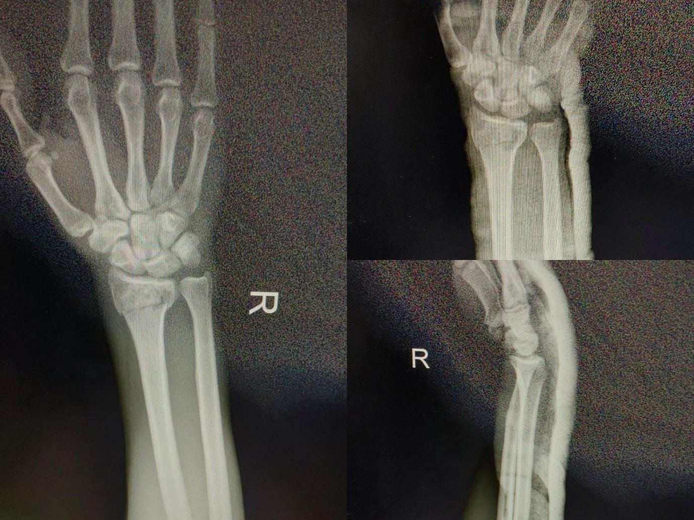
Or 28-years male labourer presented to our hospital with complaints of Pain and swelling over right wrist for one day, he had sustained trauma due to fall on open hand. On clinical examination patient had swelling, tenderness and crepitus over right wrist joint .Radiographs (Figure 1) were done which showed patient had distal radius fracture with intra articular extension fracture was minimally displaced , as Primary care his fracture was reduced with manual traction and below elbow slab was applied (Figure 1), considering the patient was right handed and the fracture was intra articular extended definite management deemed necessary for this patient , all the necessary preoperative tests were done and patient was planned for closed reduction external fixation with K-wire of distal radius fracture.

Figure 1: radiographs done at the time of presentation.
Surgery
Patient was taken in operation theatre, after two days swelling had reduced Covid swab was done for the patient, which was negative, patient was taken under All septic precautions in supine position with outstretched hand for anaesthesia supraclavicular and axillary block was given. under Image intensification fracture was examined and it was reduced with manual traction then 3 K wires were planned, first was inserted from styloid between the extensor carpi radialis and extensor carpi brevis tendons (Figure 2) and another was inserted just posterior and medial to the initial k-wire sparing the tendons and then the final k-wire was inserted from radial aspect of styloid towards ulna, holding the intra-articular fragments , this wire served the purpose of providing juxta-articular stability and compressing the fragments to little extent, surgery was completed in short period , there was minimal soft tissue damage, patient was given below elbow cast, surgery was uneventful.

Figure 2: Intra-operative Images.

Figure 3: Application of cast in immediate post-operative period for immobilisation.
Discussion
Unstable, intraarticular distal radius fractures are complicated and challenging for the surgeon. The ideal method of treatment is still an enigma. Kirschner wires are useful for provisional fixation of communicated fracture they assist in acute accurate placement of fragments and implants, especially the plates and screws, multiple wires may be inserted without any additional trauma [4]. Per-cutaneous pinning is an effective technique in maintaining the reduction of unstable extra articular fracture or an unstable moderately displaced intra articular fracture, it is also useful in obtaining and maintaining the reduction of grossly displaced intra articular fragments and is frequently used in combination with external fixation relative Contra-indications to percutaneous pinning include osteoporosis and severe comminution, planning is essential while inserting Kirschner wires for provisional fixation, pins must be introduced in such a way that do not obstruct the final placement of the definitive implants4,5.
Biomechanical studies of pin fixation support the use of minimum three pins for fixation of distal radial fragment, although for pin configuration to provide the greatest stability this may be utilised selectively as the patient is subjected to additional risk, the pins that cross radial fracture provide greater stability, the styloid radial pin is safe and avoids damage to the distal radioulnar joint [5].
In 1976, Kapandji first reported a technique of intrafocal pinning for extra-articular dorsally displaced distal radius fractures, using two conventional Kirschner wires [6]. He then made several technical modifications, and sequentially recommended the use of fully threaded pins, partially threaded pins with additional end cup, and finally the special so-called “Arum” pins. The aim was to improve the sustained buttress reduction effect of the pins, and to minimize pin related complications in terms of migration and soft tissue problems.
In 1987, he wrote a personal testimony after 10- year experience with his procedure, and acknowledged that the ideal indication for intrafocal pinning is the “simple” extra-articular Colles fracture without intra-articular extension or dorso-ulnar “third fragment”; only extra-articular fractures with minimal dorsal comminution and good bone quality may also benefit from intrafocal pinning [7].
In our case , patient being labourer belonging to lower middle class , he was selected for this procedure , though there is a lot of debate on whether, open reduction and internal fixation with volar plating would have suited the patient more but considering the need of a second surgery for implant removal, and a subset of complications which could case weakened wrist grip, and cost of second surgery and need for hospitalisation was very cumbersome for this patient ,though array of literature will uphold volar plating in this patient but with our case we can confidently say that intra-articular fracture with minimal displacement can be managed with closed reduction and k-wire though our excellent outcome in this case can be easily reproduced, careful patient selection is of utmost importance.

Figure 4: radiographs done at 8 weeks interval showing union at fracture site.
Conclusion
The closed reduction and percutaneous K-wire fixation is least invasive, safer, and functionally effective method for treating AO/ATP type A2, A3, B2 & B3 distal radius fractures. Our case had favourable outcome. Great care should be taken in every case to choose the treatment that suits the real functional needs of the patient rather than automatically pursuing a perfect radiological result using over aggressive treatment.
References
- Makhni EC, Taghinia A, Ewald T, Zurakowski D, Day CS. Comminution of the dorsal metaphysis and its effects on the radiographic outcomes of distal radius fractures. J Hand Surg Eur. 2010; 35(8): 652-658.
- Ipaktchi K, Livermore M, Lyons C, Banegas R. Current concepts in the treatment of distal radial fractures. Orthopedics 2013; 36: 778-784.
- Kapandji AI. Osteosyntheses par double embrochage intrafocal. Traitement fonctionnel des fractures non articulaires de l'extrémité inférieure du radius. Ann Chirr 1976; 30: 903-908.
- Kapandji AI. Ostéosynthèse par brochage intrafocal de type “Arum” des fractures récentes du radius distal chez l’adulte. In: Fractures du radius distal de l’adulte. Cahiers d’enseignement de la SOFCOT. Nº 67. Sous la direction de Allieu Y. Paris: Expansion Scientifique Publications, 1998: pp 67-83.
- Kapandji A. L'embrochage intra-focal des fractures de l'extrémité inférieure du radius dix ans après. Ann Chir Main 1987; 6: 57-63.
- Chatla Srinivas, Kali Vara Prasad Vadlamani, Moorthy GVS, Satish P, Narasimha Rao T, Vamshi. “Functional Outcome of Unstable Distal Radius Fractures - Treated by Closed Reduction and Percutaneous K-Wire Fixation”. Journal of Evolution of Medical and Dental Sciences 2015; 4(86): 14989-14997, DOI: 10.14260/jemds/2015/2129.
- Zemel NP. The prevention and treatment of complications from fractures of the distal radius and ulna. Hand Clin 1987; 3: 1-11.

