Down-Klinefelter Syndrome – A Rare Case of Double Aneuploidy in a Pakistani Child
Rafia Mahmood*, Saima Saad, Asad Mahmood and Sadia Ali
Department of Pathology, Armed Forces Institute of Pathology, Rawalpindi, Pakistan
Received Date: 18/08/2021; Published Date: 09/09/2021
*Corresponding author: Rafia Mahmood, Department of Pathology, Armed Forces Institute of Pathology, Rawalpindi, Pakistan
Abstract
Chromosomal abnormalities may result from a change in the chromosome number. These abnormalities may involve the autosomal chromosomes or the sex chromosomes. These may result in spontaneous abortions but major chromosomal abnormalities have been identified in live births. These individuals are usually identified early in life as they suffer from a number of congenital anomalies. Down syndrome and Klinefelter syndrome are most prevalent among autosomal and sex-linked chromosomal abnormalities, respectively. However, their occurrence together is a rare phenomenon. Few cases have been reported from around the world. These patients may present with variable clinical features, of which congenital heart disease is a common presentation. Karyotyping is a definitive diagnostic modality. Here we report a rare case of double aneuploidy, Down-Klinefelter syndrome, the first case in our population. This 13-month baby with characteristic somatic abnormalities and congenital heart disease presented for cytogenetic analysis, which showed karyotype to be 48, XXY, +21. Parents and siblings were found to have a normal karyotype.
Introduction
Congenital anomalies are an important cause of infant and childhood mortality [1]. In those who have aneuploidies compatible with life, these cause long term illness and disability and a poor quality of life [2]. These disorders lead to significant social, psychological and financial impacts on the families [3]. Congenital anomalies result from numerical and structural chromosomal aberrations [4]. Of the numerical abnormalities, aneupoidies are more commonly seen [5]. Aneuploidy is an abnormal number of one or more chromosomes [6]. These may include autosomal chromosomes, as in trisomy 21 (Down syndrome), or the sex chromosomes, like 47, XXY (Klinefelter Syndrome) [1]. These disorders individually are common causes of aneuploidy, but the incidence of both disorders in the same individual (double aneuploidy) is a rare occurrence [5].
These individuals may be suspected clinically as they may have characteristic dysmorphic features, developmental delay, intellectual disability and congenital anomalies [7]. Ultrasonography and echocardiography may assist [8]. However, definite diagnosis for exact characterization of the disorder is done by cytogenetic analysis [4]. Conventional karyotyping can effectively diagnose these disorders in most cases [6]. Prenatal diagnosis is also possible in highly suspected cases [9]. Other diagnostic modalities include fluorescent in situ hybridization, using probes specific for the suspected chromosomal abnormality and advanced techniques like array comparative genomic hybridization which is only available at limited centers and is expensive [6].
Case History
A 13-month boy was referred to our institute for cytogenetic analysis for clinically suspected Down syndrome. He was born to consanguineous parents and is the youngest of three siblings. His mother was 30 years and father were 31 years old at the time of his birth. During pregnancy, ultrasonographic examination revealed polyhydramnios. He was born full term by normal spontaneous vaginal delivery. At birth, he had an APGAR score of 10 after 1 minute. Birth weight was 2.9 kg. He developed peripheral cyanosis in the initial hours of his life. He was given oxygen inhalation and referred to cardiac center for further evaluation.
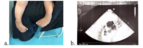
On physical examination, he had a normal height. He had Mongolian facies, slanted eyes, low set ears, thick protruding tongue and a flat occiput. Examination of his limbs revealed bow legs (varus deformity) and increased gap between his first and second toe. He had penile hypotrophy with well-developed scrotum. The child continued to have cyanotic spells, especially at time of weeping. Echocardiography showed situs solitus and levocardia. However, a 10mm ASD secundum with left to right shunt and a small left to right PDA shunting was seen. He had dilated right atrium and right ventricle with signs of volume overload.
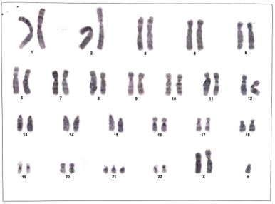
Conventional cytogenetic analysis was performed. 3 ml peripheral blood sample collected in sodium heparin was cultured in RPMI-1640. After harvesting with colchicine and treatment with KCl, slides were fixed. After banding the metaphase chromosomes using Giemsa trypsin, at least twenty metaphases were analyzed and karyotype was interpreted according to International System for Human Cytogenetic Nomenclature criteria. Analysis revealed abnormal karyotype 48, XXY, +21. Double aneuploidy – XXY and trisomy 21 was recognized in all twenty metaphases examined, consistent with diagnosis of Down-Klinefelter syndrome. There was no evidence of mosaicism. However, the standard cytogenetic methodology utilized does not routinely detect small rearrangements and low-level mosaicism, and cannot detect microdeletions. Cytogenetic analysis of parents and siblings was performed which showed normal karyotype.
Initially, he had feeding difficulties and has recurrent chest infections. The patients were counseled in detail regarding his diagnosis, further management and rehabilitation, life expectancy, possible complications and prognosis. The child is being managed by paediatric cardiologist for his cardiac problems. For his leg deformity, brace treatment is being done with regular evaluation and a plan for surgical correction. Genetic counseling with emphasis on prenatal screening for early detection of aneuplodies in future pregnancies was done.

Figure 1: a. Showing Varus deformity of lower limbs. b. Echocardiography revealed dilated RA/RV with volume overload and 10mm ASD secundum with left to right shunt.

Figure 2: Karyogram of the child showing 48, XXY, +21.
Discussion
Aneuploidy is defined as occurrence of an atypical number of chromosomes in a cell, that is, having 45 or 47 chromosomes instead of the usual 46 chromosomes [2]. These may involve the autosomal chromosomes or the sex chromosomes [1]. Double aneuploidy is a rare phenomenon. It is defined as the presence of two chromosomal abnormalities at the same time in the same individual, one involving the autosomal chromosomes while the other involving the sex chromosomes [4]. Down syndrome (Trisomy 21) is one of the most common and frequent autosomal aneuploidy with a frequency of 1 in 500 live births while Klinefelter syndrome (47 XXY) is among one of most common sex chromosome aneuploidy [7]. The exact mechanism of their occurrence together as a double aneuploidy is not clearly known. However, it may be thought that Down-Klinefelter syndrome may result from two meiotic non-disjunction events that can have same or different parental origin. The meiotic non-disjunction may occur in first or second meiotic division [6]. The incidence rate of Down-Klinefelter is about 0.098% in new born. The first case was reported by Ford et al in 1959 [10]. Since then occasional cases have been reported in different populations. This is the first case being reported in the Pakistani population.
Although Down syndrome is associated with increasing maternal age, double aneuploidy is seen in variable maternal age groups. In our patient the age of the mother was 30 years. Congenital heart disease is quite common in Down syndrome and may be seen in upto 40-50% of Down syndrome patients. However, congenital heart disease is not a feature of Klinfeleters syndrome. The frequency and type of congenital cardiac anomalies in children with Down-Klinefelter syndrome is unknown. Zheng Shen et al [6] reports the case of a child with Down Syndrome-Klinefelter Syndrome was with around to have a large ventricular septal defect (0.65 cm) and an atrial septal defect (0.55 cm) with patent ductus arteriosus (0.3 cm), pulmonary hypertension and mild tricuspid regurgitation. Gerretsen et al8 also reported a 14 month male child with double aneuploidy 48 XXY,+21 having a small atrial septal defect (secundum type) and a double aortic arch. Life expectancy of patients with Down syndrome is today up to 60 years. Lifespan is not known to be affected by Klinefelter syndrome. However, there is not enough data to comment on life expectancy of Down-Klinefelter syndrome.
Reporting of this case highlights the incidence of occurrence of this rare cytogenetic abnormality, Down-Klinefelter syndrome. It also underlines the clinical presentation of this uncommon condition. Such disorders usually remain undiagnosed. Cytogenetic analysis is a useful diagnostic modality. Early diagnosis and proper counseling of family can help improve the quality of life not only for the affected individual but can also help guide family members regarding the medical and psychosocial challenges involved.
References
- Akbas E, Soylemez F, Savasoglu K, Halliogluand O, Balci S. A male case with double aneuploidy (48, XXY, +21). Genet Couns.2008; 19(1): 59-63.
- Balwan WK, Kumar P, Raina TR, Gapta S. Double trisomy with 48, XXX+21 karyotype in a Down’s syndrome child from Jammu and Kashmir, India. Journal of Genetics, 2008; 87(3):pp 257–259.
- Suhair S, Montaha MS, MSc Ali AH, Nazmi RK, Double Trisomy 48, XXY, +21 in a Child With Phenotypic Features of Down Syndrome,Laboratory Medicine, 2009; 40(4): 215–218.
- Jeanty C, Turner C. Prenatal diagnosis of double aneuploidy, 48, XXY, +21, and review of the literature. J Ultrasound Med. 2009; 28(05): 673–681.
- Ford CE, Jones KW, Miller OJ, et al. The chromosomes in a patient showing both mongolism and the Klinefelter syndrome Lancet19591(7075): 709–710.
- Sanchez L, Petersen I, MBinkert MB, etal. A 48, XXY, +21 Down syndrome patient with additional paternal X and maternal 21. Hum Genet, 1991; 87: 54–56.
- Karaman A, Kabalar E. Double aneuploidy in a Turkish child: Down-Klinefelter syndrome. Congenit Anom (Kyoto). 2008; 48(1): 45-47. doi: 10.1111/j.1741-4520.2007.00174.x. Review. Erratum in: Congenit Anom (Kyoto). 2008; 48(2): 101.
- Rodrigues MA, Morgade LF, Dias LFA, Moreira RV, Maia PD, Sales AFH, et al. Down-Klinefelter syndrome (48, XXY, +21) in a neonate associated with congenital heart disease. Genet Mol Res, 2017; 16(3). doi: 10.4238/gmr16039780
- X Shu, C Zou, and Z Shen, Double Aneuploidy 48, XXY, +21 Associated with a Congenital Heart Defect in a Neonate, Balkan J Med Genet. 2013; 16(2): 85–90. doi: 10.2478/bjmg-2013-0038.
- Gerretsen MF, Peelen W, Rammeloo LA, Koolbergen DR, Hruda J. Double aortic arch with double aneuploidy―rare anomaly in combined Down and Klinefelter syndrome. Eur J Pediatr, 2009; 168: 1479-1481.

