Metastatic Small Cell Lung Cancer: The Role of Multidisciplinary Teams in Patient Care
Abdulsatar J Mathkhor1, Abdulnasser H Abdullah2 and Amer S. Khudhairy3
1Higher Diploma in Rheumatology and Medical Rehabilitation, Rheumatology unit in Basrah Teaching Hospital, Basrah, Iraq
2Higher Diploma in Rheumatology and Medical Rehabilitation, Rheumatology unit in Alsader Teaching Hospital, Basrah.Iraq
3Higher Diploma in Rheumatology and Medical Rehabilitation, Rheumatology unit in Alfayhaa Teaching Hospital, Basrah. Iraq
Received Date: 21/07/2021; Published Date: 25/08/2021
*Corresponding author: Abdulsatar J Mathkhor, Higher Diploma in Rheumatology and Medical Rehabilitation, Rheumatology unit in Basrah Teaching Hospital, Basrah, Iraq
Abstract
COVID-19, the disease caused by SARS-CoV-2, usually presented with pneumonia with hypoxia but can have several other manifestations and associated with different sequelae. Reactive arthritis (ReA) is sterile inflammatory arthritis, with predominant involvement of the the lower extremities. It usually occurs 1–3 weeks after a urogenital or gastrointestinal infection. An association has also been reported with bacterial and viral respiratory infections. Herein, we present 42-year-old man hospitalized following COVID-19 infection and was discharged after 10 days. After 2 weeks, he presented to the outpatient clinic complaining of severe right knee pain. After complete investigations, ultrasound, and plain X-ray, he was diagnosed with reactive arthritis. Investigations ruled out microorganisms responsible for reactive arthritis before the implication of COVID-19 infection. Nonsteroidal Anti-Inflammatory Drug (NSAID)s for 2 weeks, and intraarticular corticosteroid injection resulted in complete improvement. ReA can be autoimmune reaction sequelae after infection with COVID-19.
Keywords: COVID-19; Reactive arthritis; Spondyloarthritis
Introduction
Reactive Arthritis (ReA), a subtype of Spondyloarthritis (SpA), is sterile inflammatory arthritis causing asymmetric monoarthritis or oligoarthritis, usually of the lower limbs. It occurs 1–3 weeks after a sexually transmitted or gastrointestinal infection [1]. Chlamydia trachomatis, Campylobacter, Shigella, Salmonella, and Yersinia are the common important speices of bacteria that can cause ReA [2]. ReA also reported after infection with Streptococcus pneumonia, Staphylococcus aureus, Chlamydia pneumoniae, viral infections, and some cases were reported after COVID-19 infection [3–8]. We here report a case of ReA after COVID-19 infection.
Case Presentation
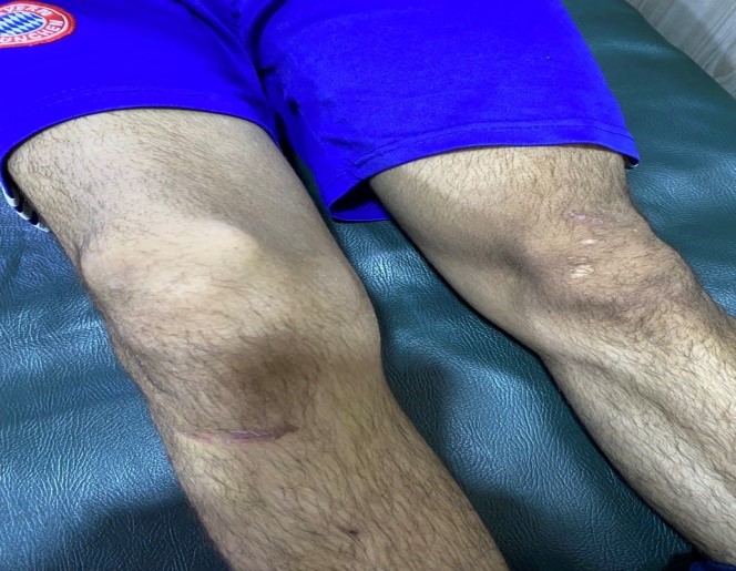
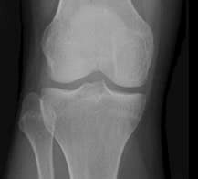
A 35-year-old man developed bone pain, dyspnoea, fatigue, and fever up to 39°C. Throat swab test for SARS-CoV- 2 infection, was positive. Additionally, CT images showed ground-glass opacity typical of viral pneumonia. Owing to the persistent fever and increasing severity of pulmonary symptoms, he was admitted to the COVID-19 ward in Basrah Teaching Hospital. After 10 days, he gradually improved and was discharged home. At the time of discharge, he was stable and had no fever, Erythrocyte Sedimentation Rate (ESR), and C reactive protein (CRP) levels were normal. Two weeks later, the patient presented to the rheumatology outpatient with severe pain and swelling in the right knee joint that prevented the patient from walking or even standing. There were no other systemic symptoms. Apart from COVID-19 infection, the patient was previously healthy and had no previous history of autoimmune disease or family history of autoimmune diseases. On clinical examination, the right knee was found swollen, warm, tender with limited movement (Figure 1), other joints not involved. He had no rash, conjunctivitis, preceding diarrhoea or urethritis. Body temperature was 36.8℃. Blood pressure was 130/80 mm Hg, heart rate 78 beats per minute, respiratory rate 18/minute. Investigations revealed normocytic normochromic anemia, leukocytosis 11.3 x10³/mm3, ESR 40, and CRP 6.8 mg/L. General urine examination was normal. Tests for ASO, HIV, rheumatoid factor (RF), anticyclic citrullinated peptide (anti CCP)antibody, antinuclear antibody (ANA) and HLA-B27 were negative. Arthrocentesis was performed for the right knee joint and synovial fluid analysis revealed mild inflammatory synovial fluid. Gram stain and of synovial fluid culture was also negative. Plain X-rays of his knee showed no periaricular osteopenia or erosive changes (Figure 2). He was diagnosed with ReA. The patient was improved after treatment with NSAIDs and intra-articular corticosteroid injection.

Figure 1: Right knee swelling.

Figure 2: Normal plain X-ray.
Discussion
Reactive arthritis is a common type of spondyloarthritis that manifests as mono- or oligoarthritis, typically with asymmetrical joint involvement following an infection with urogenital or gastrointestinal microorganisms [9,10]. The definition and diagnosis of ReA are based on a diagnostic criterion [1]. ReA following HIV infection, dengue and chikungunya viruses also was reported [11–13]. HLA-B27 and family history of spondyloarthritis apparently associated with an increased risk of developing reactive arthritis [14]. In our case, arthritis occurred two weeks after COVID-19 infection without any identifiable source of extra-articular infection and the synovial fluid cultures negative for bacteria; therefore, we strongly consider a diagnosis of clinical ReA. Few cases of ReA caused by COVID-19 have been reported. Ono K et al. were reported a ReA case following infection with SARS-COV-2 in a 57 years old man in Japan, and another case of a 73 years old man was reported by Saricaoglu EM et al. in Turkey [3,4]. Shokraee et al. described a case of ReA in the hip joint of a 58-year-old Iranian woman; in contrast, Ibtisam Jali reported ReA in hand joints in a 39-year-old Saudi Arabian woman who had no known medical illnesses apart from previous COVID-19 infection [15,16]. Lopez-Gonzales described 4 cases of acute ReA during COVID-19 illness; all these cases have rheumatologic background diseases [17]. In our case, the diagnosis of ReA is strongly supported by the classical clinical features of preceding infection (COVID-19), the absence of preceding genitourinary or gastrointestinal infection, the absence of any feature of autoimmune disease like autoantibodies, the absence of family history of autoimmune disease, and the response to NSAIDs.
Conclusion
Our case in addition to the previously described cases suggests SARS-CoV-2 infection as a potential cause in the pathogenesis of ReA. However, more studies are required to strengthen this association.
Author’s contributions
AM designed, planned the report and RS carried out the physical examinations. AK, and AH planned and carried out the laboratory tests. Finally, AM, AK, and AH participate in the writing the manuscript. All authors read and approved the final manuscript.
Patient consent for publication: Consent is obtained directly from the patient.
Ethics approval: We have obtained consent from our patient.
Acknowledgment: We kindly appreciate the patient acceptance to be enrolled in the study.
Funding disclosure: No funding was received for this manuscript
Conflicts of interest: The authors declare that there is no conflict of interest.
References
- Selmi C, Gershwin ME. Diagnosis and classification of reactive arthritis. Autoimmun Rev. 2014; 13 (4–5): 546–549.
- Carter JD, Hudson AP. Reactive Arthritis: Clinical Aspects and Medical Management. Rheum Dis Clin North Am. 2009; 35(1): 21–44.
- Ono K, Kishimoto M, Shimasaki T, Uchida H, Kurai D, Deshpande GA, et al. Reactive arthritis after COVID-19 infection. RMD Open. 2020; 6(2): 2–5.
- Saricaoglu EM, Hasanoglu I, Guner R. The first reactive arthritis case associated with COVID-19. J Med Virol. 2021; 93(1): 192–193.
- Lik YAS a, Yle HZ, Aklari LGLH. E D ‹ Töre M Ektup / L Etter To the E Ditor. 2007; 10(2): 109–112.
- Danssaert Z, Raum G, Hemtasilpa S. Reactive Arthritis in a 37-Year-Old Female With SARS-CoV2 Infection. Cureus. 2020; 12(8): 10–15.
- Sureja NP, Nandamuri D. Reactive arthritis after SARS-CoV-2 infection. Rheumatol Adv Pract. 2021; 5(1): 1–2.
- De Stefano L, Rossi S, Montecucco C, Bugatti S. Transient monoarthritis and psoriatic skin lesions following COVID-19. Ann Rheum Dis. 2020; 0(0): 1–2.
- Yu D, Lories R, Inman RD. Pathogenesis of Ankylosing Spondylitis and Reactive Arthritis. Kelley’s Textb Rheumatol. 2013; 1193–201.
- Pennisi M, Perdue J, Roulston T, Nicholas J, Schmidt E, Rolfs J. An overview of reactive arthritis. J Am Acad Physician Assist. 2019; 32(7): 25–28.
- Kishimoto M, Lee MJ, Mor A, Abeles AM, Solomon G, Pillinger MH. Syphilis mimicking Reiter’s syndrome in an HIV-positive patient. Am J Med Sci. 2006; 332(2): 90–92.
- Rich E, Hook EW, Alarcón GS, Moreland LW. Reactive arthritis in patients attending an urban sexually transmitted diseases clinic. Arthritis Rheum. 1996; 39(7): 1172–1177.
- Vassilopoulos D, Calabrese LH. Virally associated arthritis 2008: Clinical, epidemiologic, and pathophysiologic considerations. Arthritis Res Ther. 2008; 10(5): 1–8.
- Wendling D, Prati C, Chouk M, Verhoeven F. Reactive Arthritis: Treatment Challenges and Future Perspectives. Curr Rheumatol Rep. 2020; 22(7): 1–7.
- Shokraee K, Moradi S, Eftekhari T, Shajari R, Masoumi M. Reactive arthritis in the right hip following COVID-19 infection: a case report. Trop Dis Travel Med Vaccines. 2021; 7(1): 1–4.
- Jali I. Reactive Arthritis After COVID-19 Infection. Cureus. 2020; 12(11): 11–13.
- López-González MDC, Peral-Garrido ML, Calabuig I, Tovar-Sugrañes E, Jovani V, Bernabeu P, et al. Case series of acute arthritis during COVID-19 admission. Ann Rheum Dis. 2021; 80(4): 1–2.

