Testicular Teratoma with Tense Hydrocele in Newborn a Rare Presentation: A Case Report
Mahmoud M. Osman*, Mohamed S. Shoeib, Issa M. Alkhalaf, Asmaa F. Maagouze , Yasir T. Alotabi
Neonatal intensive care units (NICU), Al Yamamah Hospital, Riyadh, Saudi Arabia
Department of Radiology, Al Yamamah Hospital, Riyadh, Saudi Arabia
Received Date: 19/06/2021; Published Date: 29/06/2021
*Corresponding author: Dr. Mahmoud M. Osman, Neonatal intensive care unit, Al Yamamah Hospital, Al Manar area, Riyadh, Saudi Arabia. E-mail: osman556@hotmail.com Mobile: 00966507448117
Abstract
Testicular tumors are extremely rare in newborns. However, they should be included in the differential diagnosis of any scrotal mass. The most frequent clinical presentation is a painless scrotal swelling. Ultrasonography is the best imaging modality to study testicular tumors. Benign tumors are more frequent in prepubertal boys and malignant tumors in postpubertal. Testicular teratoma is the most common histologic type in children and is responsible for approximately 50% of prepubescent tumors. Tumor markers (alpha-fetoprotein, beta-human chorionic gonadotropin, and lactate dehydrogenase) levels contribute to the diagnosis and management of testicular masses. Herein, we present a newborn who showed a completely transilluminating right scrotal mass, that turned to be testicular cystic teratoma with hydrocele. The infant underwent a radical inguinal orchiectomy and the postoperative histological studies confirmed the diagnosis. The infant had a favorable postoperative prognosis.
Keywords: testicular teratoma, testicular neoplasms, alpha-fetoprotein, radical orchiectomy, hydrocele.
Introduction
Testicular tumors in the pediatric population are rare, and Germ Cell Tumors (GCT) are the main group of these tumors. There are two incidence peaks in children the first between 0-4 years and the second between 15–19 years, often referred to as prepubertal and postpubertal, respectively [1]. Malignant testicular tumors are more common in the postpubertal age group, and account for 12% of cancers in adolescents, compared to 4% in prepubertal children. Age is therefore described as the most important prognostic risk factor in patients with GCTs [2]. Testicular teratoma is the most frequent testicular tumor in children. Teratomas are complex tumors derived from all three germ layers (endoderm, mesoderm, and ectoderm). It is made up of several different types of tissue, such as hair, muscle, teeth, or bone. These tumors can occur in the neonatal period, however, the average age of presentation is 18 months [3]. In children with benign tumors including teratoma testicular sparing surgery has become the treatment of choice. However, radical inguinal orchiectomy is indicated in malignant tumors. The postoperative prognosis of testicular teratoma is usually excellent [4].
Case Report
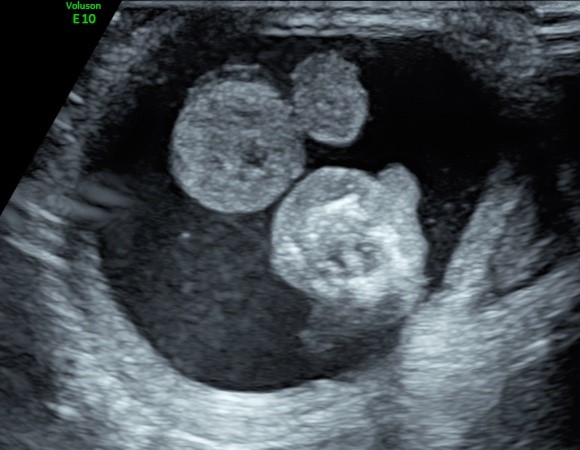
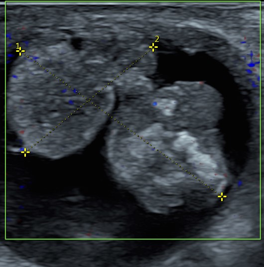
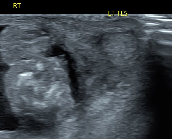
A full-term baby boy was a product of normal spontaneous vaginal delivery to a multipara 41-year-old mother who had a pregnancy complicated by severe preeclampsia. The birth weight was 3.5 kg, and the Apgar score was 8, 9, and 9 at one, five, and ten minutes, respectively. The baby received routine care and then shifted to his mother. Postnatal examination revealed a well-baby with right large cystic scrotal swelling and no other pathological findings. The baby was shifted to NICU where scrotal examination and transillumination (Figure 1) revealed a tense right hydrocele with a firm testicular mass. The left testicle was normal in position and size. Pediatric surgery consultation was done. Urgent scrotal ultrasound with color Doppler study (Figure 2 (A and B)) showed a right large cystic lesion with heterogeneous echogenicity mass, containing foci of shadowing calcifications, and soft tissue component involving the right testis. The mass measured about 2.7x1.8 cm with minimal vascularity. There was a tense right hydrocele. The left testis was normal in size, structure, and vascularity (Figure 3). There was no evidence of lymph node enlargement. The basic laboratory investigations were satisfactory, including complete blood counts, renal function tests, and liver function tests. A radiograph of the chest and abdomen was normal. Abdominopelvic ultrasound scanning was also normal. Computed Tomography (CT) scan of the chest and abdomen revealed no metastases or lymph nodes enlargement, and the rest of the study was unremarkable. The relevant tumor markers showed α‑fetoprotein (AFP) (>25,000 ng/mL; newborn normal range up to 50,000 ng/mL), β‑human chorionic gonadotropin (<1.50 IU/L; normal range 0-5 IU/L), and serum lactate dehydrogenase (216 U/L; normal range, 125-220 U/L). Based on these clinical manifestations, imaging findings, and tumor marker levels, our most likely working diagnosis was testicular cystic teratoma. At the age of 5 days, the baby was transferred to a tertiary care center with pediatric oncology and urology facilities, where the diagnosis of right testicular cystic teratoma was confirmed. The patient underwent a right radical orchiectomy and the histopathological examination of the mass revealed a prepubertal mature cystic teratoma with no neoplastic components. The postoperative course was favorable and the baby was discharged home on the 4th day postoperative in good condition. He was given close follow-ups with the urologist, oncologist, and primary neonatologist. In the subsequent visits, the levels of the α‑fetoprotein (AFP) gradually dropped to the normal level of 25 ng/mL at age of 5 months. At the time of writing this article, the baby was 6-months old in good health, with normal developmental milestones, and with no evidence of tumor recurrence.

Figure 1: The transilluminating right cystic scrotal swelling.


Figure 2 A&B : The scrotal US showed a right lobulated testicular mass with heterogeneous echogenicity contains foci of calcifications; and tense hydrocele.

Figure 3:The scrotal US showed the right testicular mass and the normal left testis
Discussion
The incidence of testicular tumors in children is only 0.5– 2.0 per 100 000 and it is less in the neonatal period. Testicular tumors are classified as germ cell tumors or non–germ cell tumors. Germ cell tumors are further classified as seminomas and non-seminomatous tumors. Seminomas are the most common testicular tumors among adults. Whereas, non-seminomatous germ cell tumors are the most common testicular tumors among children and include teratomas, yolk sac tumors, and embryonal carcinomas. Non–germ cell tumors include Sertoli and Leydig cell tumors and are rare in children [5]. Testicular teratomas are the most frequent histologic type in children and are responsible for approximately 50% of prepubertal tumors. Histologically, prepubertal teratomas can consist of any combination of the three primitive embryological germ-cell layers (ectoderm, mesoderm, and endoderm). These give rise to different tissues (epithelium, cartilage, fat, bone, muscle, and neural elements), which accounts for their heterogeneous appearance on ultrasound with areas of calcification. Teratomas are poorly or mildly vascularised on color Doppler [6]. Teratomas can occur in the neonatal period, although the average age of presentation is 18 months. Yolk sac tumors are the second most common and are most presenting at an age younger than 2 years. The prognosis can be getting better when it occurs during the first year of life [7]. Testicular tumors usually manifest as painless testicular mass in an enlarged scrotum (82–90%), and less than 10% as painful mass secondary to hemorrhage or necrosis. Physical exploration and other clinical data (fever, acute pain, vomiting) can help to differentiate a testicular tumor from hydrocele, inguinal hernia, testicular torsion, or inflammatory scrotum [8]. Mature teratomas may have a cystic quality on palpation and transillumination because of cysts filled with fluid or mucous. These testicular teratomas can be confused with hydroceles, and the diagnosis of a testicular tumor will be confirmed at surgery [9]. Nevertheless, hydroceles can be associated with testicular tumors in 15–50% of cases, and a thorough examination is needed to determine the presence of other associated pathologies, such as testicular torsion, epididymitis, and inguinal hernia [6].
Ultrasonography (US) is the imaging modality of choice for studying testicular tumors, because of its low cost, wide availability, and high sensitivity for detecting lesions. A benign tumor is suggested when ultrasonography shows a mainly cystic component, well-defined borders, or normal to increased echogenicity lesion. A malignant tumor is suspected when ultrasonography shows an inhomogeneous, hypoechoic, not well-circumscribed, or diffuse infiltration lesion. However, these ultrasonographic findings may overlap [4]. Where scrotal US findings are inconclusive or non-diagnostic, Magnetic Resonance Imaging (MRI) can contribute to ascertain the diagnosis [10]. Assessment of tumor markers, including α-fetoprotein (AFP), β-human Chorionic Gonadotropin (β-hCG), and lactate dehydrogenase, is routine and aids not only in diagnosis but also in prognosis and risk stratification. α-Fetoprotein levels are typically much higher in yolk sac tumors than in teratomas. It should be noted that infants younger than 6 months show high AFP levels physiologically, which renders interpretation is more challenging [11]. Testis-Sparing Surgery (TSS) should be used in prepubertal testicular teratomas when the normal testicular tissue seems salvageable in the US and with normal tumor markers. Intraoperative frozen section examination when available can be applied to confirm the pathology of the tumor as well as to justify the conservative surgery [12]. TSS may reduce psychological and cosmetic consequences associated with radical orchiectomy and it may reduce the risk of impaired fertility. Furthermore, in both prepubertal and postpubertal patients with a solitary testis preoperatively, TSS is recommended to preserve Leydig cell function and thereby testosterone production, and to conserve any fertility potential [13]. In the absence of a reassuring frozen section, complete orchiectomy is usually the treatment of choice. In prepubertal patients, metastatic evaluation can be deferred until a histologic diagnosis of the primary tumor has been made. Adjuvant chemotherapy in malignant testicular tumors is required. Postoperative follow-up is highly recommended for these patients with physical examination, scrotal ultrasonography, and tumor markers levels [14].
Conclusion
Although neonatal testicular tumors are rare, they should be considered in the differential diagnosis and management of a newborn with a scrotal mass. The most common histologic types of testicular tumors occurring in children are teratomas and yolk sac tumors. Testicular teratomas may transilluminate and can be confused with hydroceles, accordingly scrotal ultrasound should be considered. Timely and accurate diagnosis of scrotal swelling is essential to guide treatment decisions, and ultrasonography, color Doppler, and tumor markers play a key role in this task. We reported this case to emphasize the importance of this rare condition in newborns.
Funding and Conflict of Interest
None
References
- Kusler KA, Poynter JN. International testicular cancer incidence rates in children, adolescents and young adults. Cancer Epidemiology 2018; 56: 106–111.
- Calaminus G, Schneider DT, von Schweinitz D, Jürgens H, et al. Age-Dependent Presentation and Clinical Course of 1465 Patients Aged 0 to Less than 18 Years with Ovarian or Testicular Germ Cell Tumors; Data of the MAKEI 96 Protocol. Cancers 2020; 12(611): 1-17.
- Hisamatsu E, Takagi S, Nakagawa Y, et al. Prepubertal testicular tumors: a 20-year experience with 40 cases. Int J Urol 2010; 17: 956-959.
- Sangüesa C, Veiga D, Llavador M, Serrano A. Testicular tumors in children: an approach to diagnosis and management with pathologic correlation. Insights into Imaging 2020; 11(74): 1-14
- Edward K, Setty B, Aragon I. Sonography of the Pediatric Scrotum: Emphasis on the Torsion, Trauma, and Tumors. AJR 2012; 198: 996–1003
- Ahmed HU, Arya M, Muneer A, Mustaq I, Sebire NJ. Testicular and paratesticular tumors in the prepubertal population. Lancet Oncol 2010; 11: 476-483.
- Basta A, Courtier J, Phelps A, Copp H, MacKenzie J. Scrotal Swelling in the Neonate. J Ultrasound Med 2016; 34(3): 495–505
- Illescas T, Ibba RM, Zoppi MA, Iuculano A, Contu R, Monni G. Prenatal ultrasound diagnosis of a fetal testis granulosa cell tumour. J Obstet Gynaecol 2014; 34: 96–98
- Cathy L, Clark J. Testicular teratoma presenting as a transilluminating scrotal mass. UROLOGY 2006; 67: 1293–1295
- Mittal P, Abdalla A, Chatterjee A, Baumgarten D, Harri P, Patel J, et al. Spectrum of extratesticular and testicular pathologic conditions at scrotal MR imaging. Radiographics 2018; 38: 806–830.
- Epifanio M, Baldissera M, Esteban FG, Baldisserotto M. Mature testicular teratoma in children: multifaceted tumors on ultrasound. Urology 2014; 83: 195–197.
- Romo Muñoz MI, Núñez Cerezo V, Dore Reyes M, et al. Testicular tumours in children: Indications for testis-sparing surgery. An Pediatr (Barc) 2018; 88: 253–258
- Cheng L, Albers P, Berney M, Feldman R, Daugaard G, Gilligan T, et al. Testicular cancer. Nat. Rev. Dis. Prim 2018; 4(1): 1-24.
- Patel H, Gupta M, Cheaib G, Sharma R, Zhang A, Bass B, et al. Testis-sparing surgery and scrotal violation for testicular masses suspicious for malignancy: A systematic review and meta-analysis. Urol. Oncol 2020; 38: 344–353.

