Acute renal infarction
BEN ELHEND Salah*, DOULHOUSSNE Hassan, ROUKHSI redouane, ELFIKRI Abdelghani
Departement of Radiology, 5th Military Hospital, Guelmim, Morocco
Departement of Radiology, AVICENNE Military Hospital, Marakkech, Morocco
Received Date: 15/04/2021; Published Date: 29/04/2021
*Corresponding author: BEN ELHEND Salah, Department of Radiology, 5th Military Hospital, Guelmim, Morocco. Email: salahbenel4000@gmail.com, phone: +212 677 27 70 53
Abstract
Renal infarction is an uncommon condition resulting from a sudden disruption of blood flow in the renal artery. Patients with renal infarction typically present with acute flank pain and may have hematuria. Helical Computed Tomography (CT) must include an acquisition during the corticomedullary phase of enhancement when renal arterial and venous opacification is greatest. Helical CT findings include one or more focal parenchymal defects that involve both the cortex and medulla and extend to the capsular surface of the kidney.
Keywords: Renal infarction; Computed Tomography
Introduction
Renal infarction is an uncommon condition resulting from a sudden disruption of blood flow in the renal artery. Non-specific clinical presentation mimicking other pathologic states causes a delay in the diagnosis. Causes of renal infarction include atrial fibrillation, thromboembolism, aortic dissection, fibromuscular dysplsia renal trauma and vascularity.
Case Report
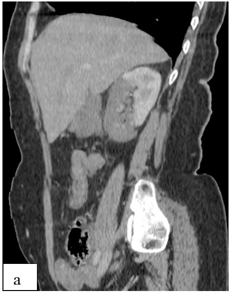
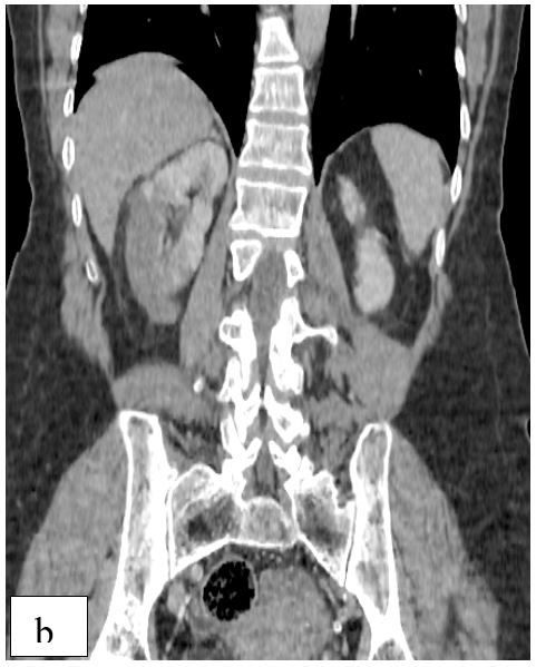
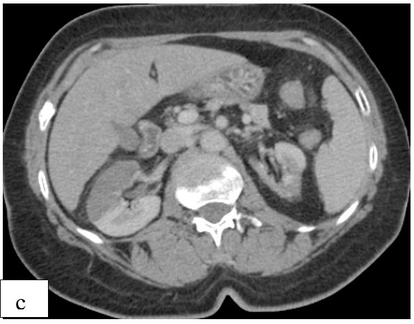
A 39-old woman was admitted for acute onset right flank pain. His has no medical history of cardiopathy. Abdominal examination revealed no defense or rebound tenderness. Vital signs as well as systemic and laboratory findings were normal, except for the elevated levels of creatinine, WBC and LDH. Urinalysis was negative for infection. Abdominal ultrasonography revealed normal findings, and doppler ultrasonography revealed no flow in the right inferior segmental renal artery. Furthermore, abdominal computed tomography with contrast revealed a hypodense area in the right kidney involving the anterolateral component of the upper and middle zones in addition to the entire lower pole.


Figure: a, b, c: Abdominal computed tomography with contrast showing a hypodense area in the right kidney involving the anterolateral component of the upper and middle zones and the entire lower pole.
Discussion
Renal infarction results from interruption of the normal blood supply to part of, or to the whole kidney. Patients with renal infarction typically present with acute flank pain and may have hematuria. The purpose of imaging and to distinguish a renal infarction from pyelonephritis or tumor. Acute infarction will appear as an absence of perfusion on color Doppler examination [1]. Helical Computed Tomography (CT) must include an acquisition during the corticomedullary phase of enhancement when renal arterial and venous opacification is greatest. Helical CT findings include one or more focal parenchymal defects that involve both the cortex and medulla and extend to the capsular surface of the kidney. Segmental infarcts of the anterior or posterior renal arteries demonstrate a characteristic appearance at CT [2]. An angiographic study of the renal vasculature is recommended in order to evaluate the presence of arterial abnormalities and localize the site of embolization or thrombosis [3].
Conclusion
Renal infarction is an uncommon condition and can lead to renal failure. The purpose of imaging and to distinguish a renal infarction from pyelonephritis or tumor.
Conflicts of Interest
The authors report no conflicts of interest.
References
- Bluth EI. Ultrasound, 2Rev Ed edition. New York: Thieme Publishing Group, 2007.
- Urban BA et Fishman EK. « Tailored Helical CT Evaluation of Acute Abdomen: RadioGraphics, 2000; 20(3): p. 725‑749.
- Mesiano et al., « Acute renal infarction: a single center experience », J. Nephrol., 2017; 30(1): p. 103‑107.

