Left Ventricle Pseudo Aneurysm in an Elderly Patient with Severe Coronary Artery Stenosis and Mitral Regurgitation
Sayarh S*, Bouazaze M, Mouine N, Asfalou I, Lakhal Z, Benyass A
Department of Medicine, Mohammed V University of Rabat, Morocco.
Received Date: 03/03/2021; Published Date: 24/03/2021
*Corresponding author: Sayarh S, Department of Medicine, Mohammed V University of Rabat, Morocco. Email:salma.s2110@gmail.com.
Introduction
Left ventricular pseudoaneurysms form when cardiac rupture is contained with adherent pericardium or scar tissue [1]. Thus, unlike a true left ventricular aneurysm, a left ventricular pseudoaneurysm contains only pericardial and fibrous elements in its wall [2]. Free intrapericardial rupture usually results in cardiac tamponade and death [3]. Less frequently, cardiac rupture is contained and left ventricular pseudoaneurysm formation occurs. Although left ventricular pseudoaneurysm is clinically uncommon, diagnosis is difficult and rupture often leads to death.
Case Report
We present a case of a 76year old man without cardiovascular risk admitted for evaluation of important shortness of breath at effort with chest pain. On examination, the temperature and vital signs were normal. Cardiac auscultation revealed a mitral regurgitation murmur.
The EKG showed a regular sinus rhythm with negative T waves in inferior and pointed T waves in anterior territory
A chest roentgenogram showed mild cardiomegaly
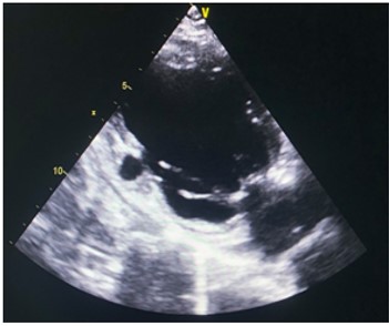
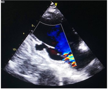
Transthoracic echocardiogram found an aspect of ischemic heart disease with undiluted and non-hypertrophied LV with akinesis of the inferior wall of the left ventricle with a left ventricular ejection fraction of 60 % complicated by a false aneurysm measuring 16/15 mm and significant is chemical mitral regurgitation by partial cord rupture. (Figure 1 and 2)
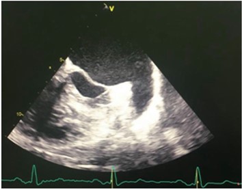
Trans esophageal ultrasound was performed showing severe ischemic mitral insufficiency by partial cord rupture with appearance of prolapse of segments A2 A3 (Figure 3)
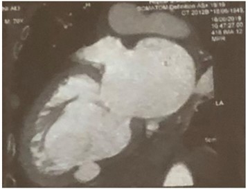
A heart CT scan with multiple reconstructions revealed an ischemic heart disease with necrosis and aneurysm of posterior LV wall. (Figure 4)
Coronary angiography showed a severe coronary artery stenosis of three vessels: ostium of the first diagonal branch, ostium of the second marginal branch and the right coronary artery
The therapeutic decision was based on a mitral repair, a bypass of the right coronary artery and the surgical cure of his anverysm because of the high risk of rupture.
In the end, the patient went out on medical treatment after refusing surgery.

Figure 1: Transthoracic echocardiogram showing a false aneurysm measuring 16/15 mm.

Figure 2: Transthoracic echocardiogram revealing significant is chemical mitral regurgitation by partial cord rupture.

Figure 3: Trans esophageal ultrasound showing a false aneurysm.

Discussion
LV pseudo-aneurysm is an uncommon but critical condition. It forms when a cardiac rupture is contained by adherent pericardium or scar tissue without endocardium or myocardium (4). There are various etiologies of LV pseudoaneurysm formation, including myocardial infarction (MI), cardiac surgery, intervention, endocarditis, and chest trauma. Frances et al. reported that 55% of cases were due to MI, and of which 49% were inferior MI (4). Meng et al. reported that the most common location of LV pseudoaneurysm formation was the posterior LV (5). Inflammatory reactions and pericardium adhesion mainly occur in the posterior wall of the LV, because patients are usually in a recumbent position after myocardial infarction (4). In the present case, pseudo-aneurysm occurred after inferior myocardial infarction.
The pseudoaneurysm is a deleterious reaction process that begins within hours of follow transmural myocardial infarction and alter the geometry of the ventricle and its wall thickness. This remodeling not only interests the infarcted zone but also the normal cardiac segments by promoting ventricular dilation according to Franck Starling's law. In the infarcted zone, systolic expansion occurs which is defined as an acute dilation and thinning from the infarct zone. This can lead to complications mechanical by rupture of the free wall of the LV or stabilization and chronic fibrosis.
Wall rupture in the acute phase can be either complete resulting in tamponade and death or incomplete forming a pseudoaneurysm as in the case of our patient. The wall of the pseudo aneurysm is then formed only from pericardial and epicardial tissue. Unlike the true aneurysm, the main differential diagnosis, the wall of which keeps all its constituents. The risk of rupture of the pseudo aneurysm is in consequence greater than that of the true aneurysm. The pseudo aneurysm can remain asymptomatic for a long time or lead to a heart failure [6].
Cardiac arrhythmias are frequently noted and can reveal the disease [7]. The aneurysmal thrombosis is a possible complication [6], with a low embolic risk. The major progressive risk is spontaneous rupture with sudden death by tamponade [6].
Clinically, a double apex beat should be researched [8]. The aneurysmal deformation of the left inferior arch on the chest X-ray is a frequent image in the case of a false left ventricular aneurysm [9], also noted in our patient.
Doppler echocardiography is the first-line test [9]. It makes it possible to make the diagnosis with certainty and to eliminate a real aneurysm. Unlike true aneurysms, which have a wall resistant fibrosis and a wider neck, the pseudo aneurysms initially consist of loose tissue, a narrower neck [10,11]. Our case presented a narrower neck at 16 mm
Cardiac CT and magnetic resonance imaging (MRI) can help distinguish the two forms and better study the specifications and location of the pseudoaneurysm [12].
After the diagnosis of LV pseudo-aneurysm is made,treatment options should be discussed.In most of cases , surgical repair is required, because of the high risk of rupture.
A previous report noted that the mortality of LV pseudo-aneurysm is considered to be higher after conservative therapy, so it should be actively treated with surgery [13] which is typically based on patch repair under extracorporeal circulation (ECC) by median sternotomy.
On the other hand, some retrospective studies have reported that cases involving chronic small LV pseudo-aneurysms of < 3cm in size or patients with high surgical risk can be managed conservatively [14].
However, rapidly progressive LV pseudo-aneurysm has also been reported [15] even in the case of conservative therapy. Periodic follow up echocardiography is mandatory.
Conclusion
Pseudo aneurysm remains a difficult diagnosis because of the symptoms, Echocardiography is used to diagnosis, and make therapeutic decision. Cardiac CT and magnetic resonance imaging (MRI) can help to determine the characteristics. Surgical indication is formal in order to prevent fetal evolution towards complete rupture.
References
- Dachman AH, Spindola-Franco H, Solomon N. Left ventricular pseudoaneurysm. Its recognition and significance. JAMA 1981; 246: 1951–1953.
- Vlodaver Z, Coe JI, Edwards JE. True and false left ventricular aneurysms. Propensity for the latter to rupture. Circulation 1975; 51: 567–572.
- Van Tassel RA, Edwards JE. Rupture of heart complicating myocardial infarction. Analysis of 40 cases including nine examples of left ventricular false aneurysm. Chest 1972; 61: 104–116
- Frances C, Romero A, Grady D. Left ventricular pseudoaneurysm. J Am Coll Cardiol 1998; 32: 557-561.
- Meng X, Yang YK, Yang KQ, et al. Clinical characteristics and outcomes of left ventricular pseudoaneurysm: A retrospective study in a single-center of China. Medicine (Baltimore) 2017 ; 96: e6793.
- Guihaire J, Verhoye JP, Flecher E, Leguerrier A. Faux anévrismes du ventricule gauche après vidéothoracoscopie : à propos d’un cas. Chir Thorac Cardiovasc 2009; 13: 120–123.
- Paraskevaidis S, Stavropoulos G, Vassilikos V, Chatzizisis YS, Polymeropoulos K, Ziakas A. Idiopathic left ventricular aneurysm causing ventricular tachycardia with 1:1 ventriculo-atrial conduction and intermittent Wenckebach block. Cardiovasc Med J 2009; 3: 105–109.
- 8.Gülera A, Uc¸ ak A, Basaran M, Ozen Y, Us MH, Yilmaz AT. Pulsatile thoracal mass: a rare case of large left ventricular pseudoaneurysm. UlusTravma Acil Cerrahi Derg 2009; 15(2): 198–200.
- 9.Ndiaye MB, Ba FG, Bodian M, Diao M, Kane AD, Sarr SA, et al. Pseudoaneurysm of the left ventricle in young patients: propos of three cases. J Ancard 2013.
- Mackenzie JW, Lemole GM. Pseudoaneurysm of the left ventricle. Tex Heart Inst J 1994;21(4):296-301.
- Prêtre R, Linka A, Jenni R, Turina MI. Surgical treatment of acquired left ventricular pseudoaneurysms. Ann Thor Surg 2000;70(2):553-7.
- Ando S, Kadokami T, Momil H et al. Left ventricular false-pseudo and pseudo aneurysm: serial observations by cardiac magnetic resonance imaging. Intern Med 2007; 46: 181-185.
- Bisoyi S, Dash AK, Nayak D, Sahoo S, Mohapatra R. Left ventricular pseudoaneurysm versus aneurysm a diagnosis dilemma. Ann Card Anaesth 2016; 19: 169-172.
- Alapati L, Chitwood WR, Cahill J, Mehra S, Movahed A. Left ventricular pseudoaneurysm: A case report and review of the literature. World J Clin Cases 2014; 2: 90-93.
- Shimono H, Kajiya T, Atsuchi Y, Atsuchi N, Ohishi M. Giant left ventricular pseudo-aneurysm after posterior myocardial infarction. Eur Heart J 2018; 39: 3479.

