Craniosynostosis in Infant with Deletion of Chromosome 4
Ioannis Drikos*
Biomedical Department, West Attica University, Athens, Hellas
Received Date: 28/01/2021; Published Date: 09/02/2021
*Corresponding author: Ioannis Drikos* Biomedical Department, West Attica University, Athens, Hellas
Abstract
Introduction: Craniosynostosis demonstrated by early fusion of one or more of the cranial sutures earlier than expected. About 8% of patients have a family history or syndromes, while the rest reported cases occur sporadically.
The inheritance of familial cases of craniosynostosis is usually autosomal dominant, affecting the action of fibroblast growth factor receptor.
Clinical case: At this study we present a case of a 4-month infant, without pediatric follow-up came to our pediatric unit with reduced feeding, vomiting and anxiety. Clinical examination and neurological assessment were normal, no signs of dehydration were observed with normal cardiac and respiratory function. During the examination of the head revealed dolichocephaly and further laboratory, imaging and surgical examination was recommended. Ultrasound analysis did not reveal any abnormalities in brain development and no other congenital abnormalities were found. Examination of the karyotype revealed deletion large portion of chromosome 4 [4q35.1].
Conclusion: Craniosynostosis occurs in many different forms. Characterized by altered head shapes can allow diagnosis. It is associated in most cases with mutations in genes based on chromosome 4 and related to fibroblast activity
Keywords: Cranyosinostosis; Infant; Chromosomal deletion
Introduction
Craniosynostosis reveal early fusion of one or more of the cranial sutures. About 8% of patients referred family history or syndromes, while the rest report cases acquired seizures occur individually. Its incidence is estimated at about 1 in 2000-2500 births. It can be spontaneous, syndromic, or familial and the heritance of familial cases of craniosynostosis is usually autosomal dominant stopped the action of the fibroblast growth factor receptor. Due to the risks associated with brain development and the proper structure of the head, the treatment of craniosynostosis is surgical and is usually performed immediately after diagnosis. Early referral to specialized pediatric rehabilitation center with accompanying evaluation by pediatric neurologists can therefore prevent complications of craniosynostosis.
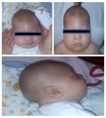
Clinical case: A 4-month infant was examined in our pediatric unit for vomiting and reduced feeding. The infant was in good general condition, lively, with a myxedematous face and oblique eyelid fissures. The neurological examination was normal and the clinical examination determined macrocephaly with possible diagnosis of craniosynostosis (Figure 1). Due to history, it was recommended to perform laboratory and imaging tests. The laboratory test revealed WBC: 12500 / μL, W: 28%, L: 59%, M: 9%, Hgb: 9.5 gr / dl, Hct: 26.9%, MCV: 80.9 fl, PLT: 570000 / μL , TKE: mm, CRP: 3.12 mg / L, Glucose: 109 mg / dl, Urea: 15 mg / dl, Creatinine: 0.2 mg / dl, SGOT: 51 IU / L, SGPT: 44 IU / L , Na: 140 mmol / L, K 4,4: mmol / L, Cl: 100 mmol / L, Total Hall: 3.51, Direct Hall: 0.79, P: 5.6, Mg: 2.07, Total Albums: 5.10 albumin: 3.89 globin’s: 1.21 PT = 12.4 sec, INR = 0.96, aPTT = 30.6, fibrinogen = 235. Ultrasound examination of the heart, upper and lower abdomen and brain ultrasound did not reveal any abnormalities. Examination of the karyotype revealed deletion of chromosome 4 [4q35.1]. Immediate assessment by a pediatric neurosurgeon was recommended to assess craniosynostosis and treated condition surgically.
Discussion: Several syndromes have been described to be related to various facial features as well as craniosynostosis. The main complication of craniosynostosis is the appearance of hydrocephalus. According to published studies, fibroblasts appear to have crucial role with the growth factor receptor being frequently modified. The growth factor receptor has tyrosine kinase activity and is crucial in differentiation and maturation of osteoblasts [1,2].
The mode of inheritance is autosomal dominant. The sagittal suture is usually more affected and may be associated with fingertip or midline hypoplasia. Most cases refer to be spon-taneous while risk factors include low birth weight, premature birth, use of drugs by the mother and obstructive causes that can lead to hydrocephalus [3,4]. Deformities of the skull can lead to growth retardation and consequent increased Intracranial Pressure (ICP). Deformity can lead to psychological and social behavior problems. In addition to genetic factors craniosynostosis may develop in patients with restricted venous flow to the sphincter, with obstructive sleep apnea and hydrocephalus or obstruction of the fourth ventricle [5]. Craniosynostosis can lead to increased intracranial pressure at about 30% to 44% of cases. Increased intracranial pressure is usually monitored as non-invasive and may be associated with symptoms such as headache, nausea, vomiting and paralysis [6]. As already mentioned main causal factors are genetic. In 6 to 11% of children born with craniosynostosis, family history is reported such as mutations in fibroblast genes (FGFR3) based on chromosome 4 and TWIST [7, 8]. Fibroblast growth factor receptors and fibroblast growth factors regulate fetal bone growth and associated with the development of cranial sutures [7, 8]. TWIST gene reacts action of the growth factor receptor and appears to reduce FGFR function directly regulating fetal bone growth [9].In 31% of cases mutations in the FGFR3 gene leads to premature convergence of the coronary suture. Mutations in the FGFR1 and FGFR2 genes have been reported in 90% of cases of craniosynostosis-related syndromes such as Pfeiffer and Jackson-Weiss [11]. These genes are located on chromosome 4 and chromosomal mutations on that particular chromosome may be associated with craniosynostosis. Appearance of craniosynthesis may be related to differential regulation pathways and balance between the action of genes that contribute skull development [11, 12]. The treatment is surgical and the operation is generally performed after the child reaches the age of 6 months due to the reduction of the surgical risk [3, 6].

Figure 1: Craniosynostosis in an infant with deletion of chro- mosome 4.
Discussion
Craniosynostosis occurs in many different forms inherited with relevant head shapes allow diagnosis. It is most often associated with mutations in genes based on chromosome 4 and related to fibroblast activity. Early diagnosis and detection of the shape of the skull contributes effective treatment and prevention of developmental disorders and hydrocephalus.
References
- Aleck K. Craniosynostosis syndromes in the genomic era. Semin Pediatr Neurol 2004; 11(4): 256-261.
- Kimonis V, Gold JA, Hoffan TL, Panchal J, Boyadjiev SA. Genetics of craniosynostosis. Semin Pediatr Neurol 2007; 14(3): 150-161.
- Czerwinski M, Kolar JC, Fearon JA. Complex craniosynostosis. Plast Reconstr Surg 2011; 128(4): 955-961.
- Ursitti F, Fadda T, Papetti L, Pagnoni M, Nicita F, Lannetii G et al. Evaluation and management of no syndromic craniosynostosis. Acta Pediatric 2011; 100(9): 1185-94.
- Di Rocco F, Arnaud E, Renier D. Evolution in the frequency of no syndromic craniosynostosis. J Neurosurg Pediatr 2009; 4: 21- 25.
- Derderian C, Seaward, J. Syndromic Craniosynostosis. Semin Plast Surg 2012: 26: 64-75.
Stamper BD, Park SS, Beyer RP, Bammler TK, Frain FM, Mecham B et al. Suture Craniosynostosis. PLoS One 2011; 6(10): 265-557. - Marie PJ. Fibroblast growth factor signaling controlling osteoblast differentiation. Gene 2003; 316: 23-32.
- Mulliken JB, Gripp KW, Stolle CA, Steinberger D, Müller U. Molecular analysis of patients with synostotic frontal plagiocephaly (unilateral coronal synostosis. Plastic and Reconstructive Surgery. 2004; 113 (7): 1899–1909.
- Agrawal D, Steinbok P, Cochrane DD. Scaphocephaly or dolichocephaly? Journal of Neurosurgery. 2005; 102 (2 Suppl): 253–254.
- Lenton KA, Nacamuli RP, Wan DC, Helms JA, Longaker MT. Cranial suture biology. Current Topics in Developmental Biology.2005; 66: 287–328.
- Passos-Bueno MR, Serti Eacute AE, Jehee FS, Fanganiello R, Yeh E. Genetics of craniosynostosis: genes, syndromes, mutations and genotype-phenotype

