Dilated Ascending Aorta in Crohn’s Disease: Unusual Association
Larsen Clarck MOUMPALA ZINGOULA, Sara AHCHOUCH, Divine TCHILOEMBA, Meryem BENNANI,Ali CHAIB, Ilyass ASFALOU, Aatif BENYASS
Resident in Cardiology, Assigned to Non-Invasive Exploration (HMIMV), Ibn Sina University Hospital Center, Mohammed V Military Instruction Hospital, Morocco
Cardiology Specialist, Assigned to Non-Invasive Exploration (HMIMV), Mohammed V Military Instruction Hospital,Morocco
Resident in Cardiology, Assigned in Rhythmology (HMIMV), Ibn Sina University Hospital Center, Mohammed V Military Instruction Hospital, Morocco
Cardiology Specialist, Assigned in Rhythmology (HMIMV), Mohammed V Military Instruction Hospital, Morocco
Professor in Cardiology, Head of Rhythmology, Mohammed V Military Instruction Hospital, Morocco
Professor of Cardiology, Head of Non-Invasive Exploration, Mohammed V Military Instruction Hospital (HMIMV), Morocco
Professor in cardiology ,head of the cardiology center of the military hospital of Instruction Mohammed V
Received Date: 19/12/2020; Published Date: 22/01/2021
*Corresponding author: Larsen Clarck MOUMPALA ZINGOULA, Resident in Cardiology, Assigned to Non-Invasive Exploration (HMIMV), Mohammed V University of Rabat, larsenmoumpala@gmail.com, 00212684804092
Summary
The association of dilatation of the ascending post junctional aorta and Crohn's disease has not been described. Although not malformative, this dilation can be inflammatory. Usually asymptomatic, the diagnosis is discovered incidentally when an imaging test is performed for another cause. We report the unusual case of a young patient with dilating ascending aorta associated with Crohn's disease.
Keywords: Dilation of the ascending aorta; Chronic inflammatory bowel disease; Crohn's disease.
Introduction
The aneurysm of the ascending thoracic aorta is the result of an alteration of the media of the aorta which is manifested histologically by: a progressive decrease in smooth muscle cells replaced by areas of mucoid degeneration rich in vacuoles and in polysaccharides; disorganization and loss of elastic fibers in the extracellular matrix. These processes result in weakening of the aortic wall, which in turn is responsible for its dilation, the causes of which are either familial (syndromic, monogenic and congenital), inflammatory (Takayasu's disease, Horton's disease, etc.) and degenerative [1].
Crohn's Disease (CD) is one of the chronic Inflammatory Bowel Diseases (IBD) alongside ulcerative colitis of the intestine with systemic osteoarticular, coronary and cerebrovascular manifestations. It affects 140 people per 100,000 inhabitants and six women for four men. The majority of cases are diagnosed around the age of 25 [2].
We report a case of Dilatation of the Ascending Aorta Post Junctional (DAAPJ) associated with chronic Inflammatory Bowel Disease (IBD).
Clinical Case
This is a 26-year-old patient, without cardiovascular risk factors, without professional sports activity, followed in rheumatology for iterative episodes of febrile polyarthralgia accompanied by watery diarrhea which has progressed for 3 years with a negative genetic test for HLA B27.
On clinical examination, the patient was febrile (38.5 ° C) he presented a tachycardia at 105 bpm, a respiratory rate at 18 cpm, had blood pressure figures around 100/60 mmHg, the cardiovascular and pleuro examination. pulmonary is normal, his electrocardiogram is unremarkable; in biology: hyperleukocytosis (20,200 / ul) predominantly neutrophilic at 17,089 / ul and a positive inflammatory assessment with an ESR 39 mm, CRP at 364 mg / l, ferritin at 1429 ng / ml, a procalcitonin negative, negative serologies, negative blood cultures. The rest of the assessment was unremarkable, in particular the renal assessment (DFG at 122 ml / min / 1.73m2).
The chest and spine radiologies were unremarkable.
He benefits:
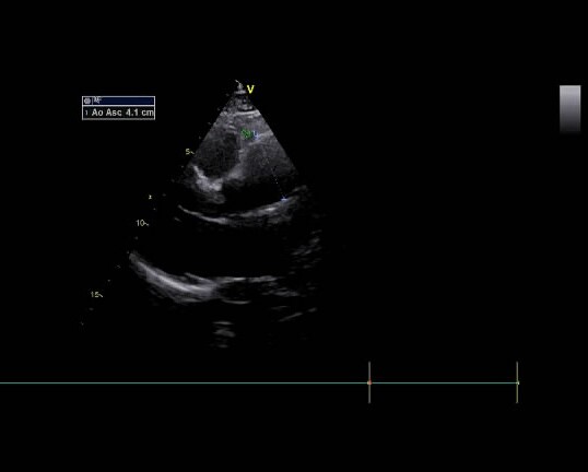
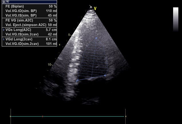
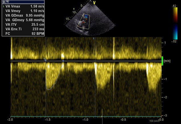
- a) a Doppler echocardiography which shows an undilated left ventricle, not hypertrophied with an ejection fraction preserved at 59%, with an ascending aorta dilated to 41 mm slightly sacciform at the anterior wall in longitudinal section (without involvement of the aortic base or sinus) (Figure 1-3).
- b) thoraco-abdominal-pelvic CT angiography showing moderate dilation of the ascending aorta of 40 mm without aneurysm (Figure 4,5).
- c) Colonoscopy: normal in appearance, including an anatomic pathological biopsy sample showing ulcerated subacute colitis which may be part of an IBD of Crohn's disease type.

Figure 1: Sacciform dilation of the post-junctional ascending aorta at 41 mm. HMIMV Non-Invasive Cardiology Exploration Service.

Figure 2: Left ventricle, undilated, left ventricular ejection fraction retained. HMIMV Non-Invasive Cardiology Service

Figure 3: Maximum velocity of aortic flow normal, without aortic leakage. HMIMV Non-Invasive Cardiology Service.

Figure 4: Dilated ascending aorta, without aneurysm, before injection of contrast product. HMIMV Radiology Department.

Figure 5: Dilated ascending aorta, without aneurysm after injection of contrast product. Radiology Department of the HMIMV.
Discussion
Given the young age of our patient, it would be wise to first rule out a congenital malformative cause.
Marfan syndrome (and related) was mentioned first. However, it combines cardiovascular and skeletal signs, lens ectopia and pneumothorax [1,3]. which is not the case in our patient. However, the Ankylosing Spondylitis-Marfan combination was described by P. Fietta and P. Manganelli in a 46-year-old patient, who had complete Marfan syndrome [4].
The aneurysm of the ascending thoracic aorta is the result of an alteration of the media of the aorta which is reflected histologically by: a progressive decrease in smooth muscle cells replaced by areas of mucoid degeneration rich in vacuoles and in polysaccharides; disorganization and loss of elastic fibers in the extracellular matrix. These processes lead to weakening of the aortic wall, which in turn is responsible for its dilation [1].
Indeed, aneurysms affecting the ascending aorta, of degenerative origin, are not linked to atherosclerosis (unlike aneurysms of the abdominal aorta and the descending thoracic aorta) but rather to a genetic part. In addition, we know that the ascending and descending thoracic aortas, separated anatomically by the ligamentum arteriosum, are two very distinct entities. This observation seems to be based in part on the difference in their embryonic origin [1].
[Figure 1-5]
The presence of inflammation or suggestive symptoms should suggest the association of dilation of the ascending aorta with inflammatory disease. In the first ranks, we find the vasculitis of the large caliber arteries: Takayasu's disease, giant cell arteritis, Behçet's disease but also spondyloarthropathies, connectivitis and finally infectious diseases such as syphilis or tuberculosis [5]. Crohn's is associated with extraintestinal manifestations and other autoimmune pathologies much more frequently than ulcerative colitis. Physiopathologically, it is a vasculitis of the mesenteric arteries which is thought to cause ischemic lesions and multiple infarctions of the intestinal wall [6,7]. Recent studies have shown that patients with IBD have a moderately increased risk of cardiovascular complications (ischemic heart disease, stroke and mesenteric ischemia), with no difference to Crohn's disease and UC [8]. To date no description associating IBD and dilation of the ascending aorta has not been reported to our knowledge.
However, in a case reported by El Kouache in 2013 of Ankylosing Spondylitis (AS) complicated by dystrophic aneurysm of the ascending aorta, would be similar to ours in an inflammatory context. AS, an inflammatory disease, can be complicated by systemic manifestations, including inflammatory cardiovascular disease, especially aortic disease. The histology of this aortic involvement, reported by El Kouache, is characterized by an inflammatory infiltrate predominantly lymphocytic which involves the first 3 centimeters of the aortic root. This results in adventitial thickening, destruction and focal necrosis of the elastic tissue of the media as well as intimal proliferation. In this observation, the histological study of the aortic wall did not identify inflammatory changes but rather cystic degeneration with dissociation of the bundles of elastic fibers observed throughout the thickness of the media. The intima was little changed. This histological description suggests dystrophic involvement comparable to that seen in Marfan's disease, but this 47-year-old patient had no other diagnostic criteria for this syndrome. This observation therefore raises the question of a possible relationship between AS and the histological lesions of Marfan [9]. However, we excluded the diagnosis of Marfan in our patient.
Ridker et al. showed, for the first time in 2003, that circulating levels of ultrasensitive C-Reactive Protein (CRP) strongly correlated with the subsequent risk of CV complications in the general population [10].
Supported
Laplace's law states that the more the tension exerted on the wall increases, the more the diameter of the aneurysm increases leading to dilation of the aorta. Thus, the kinetic energy produced during turbulence has an important effect on the distribution of pressure on the sacciform wall. It is therefore considered to be an important factor in avresysmal dilation as well as its rupture [11].
According to the recommendations of various Cardiology Societies, only recommend surgery for diameters> 50-55 mm and favor medical treatment for a diameter <40 mm [11].
Thus, we put our patient on beta blockers, because they made it possible to decrease the progression of the aortic aneurysm, according to Laplace's law by negative inotropic and chronotropic effect, while improving life expectancy, recommended class I, B [1]. As for the ARS inhibitors, our patient presents blood pressure limits between 90 and 100 systoles, hence abstention. As for the statin, it would seem that it has a beneficial effect, in particular on its ascending portion, by reducing its speed of progression linked to its anti-inflammatory action (in vivo and in vitro, statins decrease the expression of metalloproteins which are involved in the dilation process) [1].
As for his Crohn's disease, he is put on methotrexate 25 mg 1 tab / day [2].
Clinico-biological and imaging monitoring is recommended every 12 months [1].
Conclusions
Dilatation of the ascending post junctional aorta in a young patient associated with Crohn's disease remains an unusual case. However, a histological study of the ascending aorta would have confirmed at best the attribution of Crohn's disease in this dilation. As for its management, it remains medical for a diameter <40 mm and surgical if greater than 50 mm.
References
- Artigou JY, Monsuez JJ, et al. Cardiology and Vascular Diseases. French Society of Cardiology. Premium offer. Edition 2020, Elsevier Masson SAS.
- Vincent REYT. Crohn's disease. Prescription analysis, gastroenterology, Elsevier Masson 2018.
- Dupuis C, Kachaner J, Freedom RM, Payot M, Davignon A. Pediatric cardiology, 2nd edition. 1991 by Flammarion.
- Fietta P, Manganelli P. Coexist Marfan's syndrome and ankylosing spondylitis: a case report. Clin Rheumatol 2001.
- Mirault T. Rare diseases and dilation of the ascending aorta, focus. Journal of Vascular Medicine. Elsevier Masson, March 2018.
- Ben Youssef H. Arterial damage and chronic inflammatory bowel disease: series of 6 cases and review of the literature. Journal of Vascular Diseases. Elsevier Masson, 2016.
- Nguyen MC, et al. Takayasu disease associated with Crohn's disease: two identical diseases at different sites? Rev Med Switzerland 2011; 7: 1325-1328.
- Romain Altwegg. Cardiovascular complications and IBD. Montpellier University Hospital. Afa Crhon-RCH-France, 2017.
- El Kouache M, Benzidia R, Benomar M, S. Moughil. AS complicated by dystrophic aneurysm of the ascending aorta: systemic fibrillinopathy? Letter from the Rheumatologist. N ° 395, 2013.
- Ridker PM, et al. C-reactive protein, the metabolic syndrome, and risk of incident cardiovascular events: An 8-year follow-up of initially healthy American women. Circulation 2003; 107: 391-397.
- Al-Attar N, Nataf P. Dilation of the ascending aorta: when to operate? Cardiological Realities 278, 2011.

