Female Pelvis Case Report: How a Traditional Anatomy Class Can Help Medical Students?
Danilo Euclides F*, DeizeGrazielle Conceição FF and Alexandre Augusto PC
1Medical student, Universidade Federal de São Paulo, Brazil
2Assistant professor, Universidade Federal de São Paulo, Brazil
Received Date: 04/08/2020; Published Date: 18/11/2020
*Corresponding author: Danilo Euclides Fernande, Department of Medicine, Universidade Federal de São Paulo, Botucatu St. 591, 15th floor, room 153, SP, 04023-900, Brazil. E-mail: defernandes@unifesp.br
Abstract
Background: During anatomy (topographic) chair, we prepared an anatomic piece of an older female pelvis (unknown causa mortis). Here, we present an anatomic piece prepared by medical students that helped us to better understand hypertrophic bladder pathophysiology.
Methods: A traditional corpse dissection guided by an assistant professor
Results: Corpse dissection enhanced our personal experience during the medical course. It also helped us to better visualize how big a hypertrophic urinary bladder can become as the etiologic cause is not taken care.
Conclusions: Despite virtual and 3D anatomy lessons, we believe corpse dissection must remain as a teaching strategy that can help to build a new generation of surgeons as well as honor the History of Medicine.
Keywords: Anatomy; Medical Education; Teaching; Students; Graduate Medical Education
Introduction
During the anatomy (topographic) chair at our institution, we dissected and prepared an anatomic piece of an older female pelvis (unknown causa mortis). Her corpse was donated to the Universidade Federal de São Paulo to help medical students to learn clinical-surgical anatomy. As medical education becomes more and more modern, corpses dissection remains on the history of anatomy teaching and gives space to new technological strategies. Though, we believe some practical skills and surgical abilities can be learned the old-fashioned way through a traditional anatomy lesson. Here, we present an anatomic piece we prepared during our anatomy classes that helped us to better understand hypertrophic bladder pathophysiology. After analyzing the internal genital organs (hypoplastic uterus and ovaries) and the angles, we concluded this pelvis belonged to an old female person.
Diagnostic and discussion
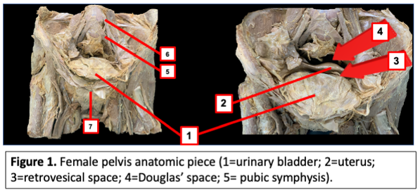
Figure 1 shows the anatomical piece the author prepared under the orientation of Dr. Alexandre Cardoso. During the dissection, we recognized urinary bladder hypertrophy which usually happens in the presence of an outlet obstruction. The most frequent causative factor is benign prostatic hyperplasia in men [1]. Among common causes of bladder outflow obstruction, in women, are [2]:
- pelvic organ prolapse
- uterine fibroid or tumor
- urethral disorders (meatal stenosis, urethral caruncle)
- luminal impaired detrusor (stone, bladder/urethral tumor, ureterocele, foreign body)
- contractility disorder (senile bladder changes, neurological diseases suchas diabetes mellitus or another peripheral neuropathy); and
- functional disorders (such as Fowler's syndrome).
Clinically, even though this is a rare condition among women, clinical investigation must ask poor urinary stream and incomplete bladder emptying. Physical examination should search for mechanical urethral obstruction. Nowadays, ultrasonography has been considered a good non-invasive strategy because of its capacity of assessing presence of residual urine volume and determination of patient’s Uroflow which must be > 15 ml/s on a voided urine volume of over 150 ml [2]. Then, if one suspect of urethral obstruction, clinical history, physical examination and ultrasound may help to diagnose it. As third-year medical students, corpse dissection enhanced our personalexperience during the medical course. It allowed us to manipulate the internal organs as far as we needed to understand its topography in relation to bone and muscle structures. It also helped us to better visualize how big a hypertrophic urinary bladder can become as the etiologic cause is not taken care. Despite our virtual and 3D anatomy lessons, we definitely believe corpse dissection must remain as a teaching strategythat can help to build a new generation of surgeons as well as honor the History of Medicine.
As recently shown, both virtual and cadaver-based anatomy class enhance first year medical students’ experience [3]. Besides, the students may be inspired by corpse dissection to easily engage on studying pathophysiology because it supports their learning processes affectively. It also augment strategies to easily memorize medical contents as well as apply their knowledge to future clinical practice.

References:
- Fusco F, Creta M, De Nunzio C, Iacovelli V, Mangiapia F, Li Marzi V, et al. Progressive bladder remodeling due to bladder outlet obstruction: a systematic review of morphological and molecular evidences in humans. BMC Urol. 9 de março de. 2018;18(1):15.
- Yande S, Joshi M. Bladder outlet obstruction in women. J -Life Health. 2011;2(1):11-7.

