An Unusual Case of Steroid Resistant IgG4-RD Affecting the Maxillary Alveolar Process
Shannon S*, Wildan T and Perry M
1Department of Oral and Maxillofacial, Northwick Park Hospital, London North West University, Healthcare NHS Trust, London
Received Date: 09/05/2020; Published Date: 26/05/2020
*Corresponding author: Sarah Shannon, Department of Oral and Maxillofacial, Northwick Park Hospital, London North West University, Healthcare NHS Trust, London, HA1 3UJ. E-mail: sarah.shannon3@nhs.net
Abstract
Immunoglobulin G4-related diseaseis an autoimmune fibro-inflammatory condition which is characterisedhistologically by marked lymphoplasmacytic infiltration, storiform fibrosis and obliterative phlebitis. Immunostaining reveals IgG4+ plasma cells. Immunoglobulin G4-related diseaseaffecting the head and neck region is becoming increasingly recognised and generally shows a good initial response to steroids. Common manifestations within the head and neck include the orbit and the salivary glands. This article presents a rare case of immunoglobulin G4-related diseaseoriginating in the alveolar process of the maxilla with orbital and maxillary sinus involvement which wasunresponsive to management with steroids.
Case Report
A 37 year old Somalian woman was referred to the Oral and Maxillo-Facial Surgery department with a 4 month history of progressive swelling of her right cheek. She had multiple presenting complaints including paresthesia and neuropathic pain affecting the V2 and V3 regions of the right trigeminal nerve. She had difficulty opening her mouth. She was not experiencing any visual issues at this initial presentation. However, she was experiencing subjective hearing impairment of the right ear. These symptoms had a gradual onset on a background of her being systemically well. She was taking only intermittent vitamin D supplements.
Examination
Examination showed a defined swelling over the right maxilla which was tender to palpation. Mouth opening was reduced. Examination of cranial nerves I, II, III, IV, VI, VIII, IX, X, XI and XIIwasunremarkable. However, testing of cranial nerve V revealed that she had reducedperception to light touch over the site of swelling. Normal sensory perception was maintained elsewhere in the V1 and V3 regions and the contralateral face. Hearing was reduced on the right side on whisper test.
Investigations
An ultrasound scan revealed an irregular, hypoechoic lesion in the soft tissues of theright cheek. The lesion was deep to the subcutaneous tissue and lacked any significant internal vascularity. A computed tomography scan and magnetic resonance imaging of the head and neck defined; A 5cm mass centred on the right maxillary alveolus and hard palate extending into the right buccal space, right masticator space and right inferior orbital fissure.Destruction of the right maxillary alveolus and hard palate, Involvement of the right infra-orbital and greater palatine nerves
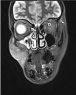
Figure 1a: Coronal view

Figure 1b: Axial view of the MRI head showing an aggressive focal lesion causing destruction of the right maxillary alveolus/hard palate and the maxillary sinus with extension to the right buccal space, right masticator space and the right inferior orbital fissure with involvement of the rightinfraorbitaland greater palatine nerves.
A tissue biopsy of the site was taken to plan treatment. This showed a storiform pattern of stromal fibrosis with an intense inflammatory infiltrate including lymphoid follicles with reactive germinal centres. Obliterativevasculitis was seen. Of note, there was an increased number of IgG4+ plasma cells on immunostaining suggesting a IgG4 fibrosclerotic lesion.
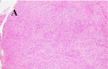
Figure 2a
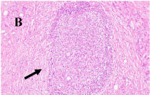
Figure 2b
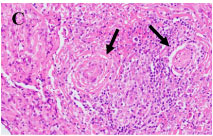
Figure 2c
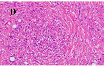
Figure 2d
Serum IgG4 levels were within normal range and IgG1 levels were elevated.Diagnosis of immunoglobulin G4-related diseasewas thus confirmed on the basis of clinical and histopathological findings outlined above.
Treatment
The patientwas urgently referred to the Rheumatology team who started her on a course of prednisolone however there was no response to management with steroids. The swelling progressed with subsequent right sided proptosis and ptosis. She was therefore started on cyclophosphamide and rituximab. This subsequentlyshowed an improvement clinically and symptomatically.
Discussion
Clinically, immunoglobulin G4-related disease presents as a tumefactive, tissue destructive lesion [1] and often mimics malignant tumours which can complicate diagnosis [2]. It can affect multiple different organs throughout the body but is becoming increasingly recognised within the head and neck. Common sites of involvement within the head and neck include the orbits and the salivary glands [3]. Immunoglobulin G4-related diseasemanifesting in the maxilla is rare. There are only three reports of the disease affecting the alveolar process of the maxilla within the literature to date [4-6]. As presented in this case report, patients with immunoglobulin G4-related maxillary disease may experience swelling, facial pain [4,6] and trismus[6]. In our case proptosis and pseudoptosisoccurred as the disease progressed and the orbit became involved. There was no diplopia and visual acuity was grossly intact. Pupillary reflexes were normal. Other possible symptoms that have been reported in patients with maxillary immunoglobulin G4-related diseaseinclude increasing mobility and loss of teeth [4] and dysphagia [6].
In any patient presenting with an expanding / invading mass urgent diagnosis is required to enable appropriate and timely treatment (surgical excision, chemo-radiotherapy or other non-surgical treatments, as in this case). Diagnosis of immunoglobulin G4-related disease is usuallybased upon a combination of clinical, histopathological and serological findings. It is characterised by the presence of an intense lymphoplasmacytic infiltration, storiform fibrosis and obliterative phlebitis on histopathological examination. Immunostaining reveals IgG4+ plasma cells. Serum levels of IgG4 may also be raised in patients with IgG4-RD. However, elevated serum levels of IgG4 are not diagnostic of the disease [7]. Evidence suggests that many as 30% of patients with biopsy confirmed immunoglobulin G4-related diseasecan have normal serum levels [8].
Currently, the first line treatment modality for immunoglobulin G4-related disease is systemic glucocorticoids. [2,9] A systematic review of the therapeutic approaches to immunoglobulin G4-related diseaseshowed glucocorticoids to have a 96% efficacy as a first line treatment [10]. This article presents a rare case of immunoglobulin G4-related disease which is non-responsive to treatment with steroids and outlines an alternative management using cyclophosphamide and rituximab. Patients treated with cyclophosphamide and concurrent steroid therapy have been shown to have a higher rate of complete remission and a lower rate of relapse in comparison to those treated with glucocorticoid monotherapy [11]. Rituximab is a third line treatment agent for immunoglobulin G4-related disease in patients who fail to respond to treatment with steroids and is used to successfully prevent further disease progression [2]. In this case report, management with a combination of these medications resulted in an improvement both clinically and symptomatically.
Conflict of Interest
Sarah Shannon, Tun Wildan and Michael Perry declare that they have no conflict of interest
References:
- Stone JH, Zen Y, Deshpande V. IgG4‐Related Disease. N Engl J Med. 2012;66:539-51.
- Lang D, Zwerina J, Pieringer H. IgG4-Related Disease: Current Challenges And Future Prospects. The Clin Risk Manag. 2016;12:189-199.
- Mulholland GB, Jeffery CC, Satija P, Côté DW. Immunoglobulin G4-Related Diseases in The Head And Neck: A Systematic Review. J Otolaryngol Head Neck Surg. 2015;44:24.
- Kouwenberg, WL, Dieleman FJ, Willems SM. Inflammatory Pseudotumour of The Alveolar Process oof The Maxilla As Clinical Manifestation Of IgG4-Related Disease: A Case Report And Literature Review. Int J Oral Maxillofac Surg. 2019;19:31406-7.
- Rodriguez Fonseca OD, Suarez JP, Dominguez ML, Fernandez Llana B, Vigil C, Martin N et al. Isolated Immunoglobulin G4-Related Disease of Nasal Septum and Maxilla: Diagnosis and Follow-up With 18F-FDG PET/CT. Clin Nucl Med. 2020;45:122-124.
- Santana IU, de Fonseca EP, Santiago MB. IgG4-Related Disease: A Multispecialty Condition. Case Rep Rheumatol. 2014;723493.
- Deshpande V, Zen Y, Chan JK, Yi EE, Sato Y, Yoshino T et al. Consensus Statement On The Pathology Of IgG4‐related disease. Mod Pathol. 2012;25:1181-92.
- Carruthers M, Khosroshahi A, Augustin T, Deshpande V, Stone JH. The Diagnostic Utility Of Serum IgG4 Concentrations In IgG4‐Related Disease. Ann Rheum Dis 2015;74:14-8.
- Khosroshahi A, Wallace ZS, Crowe JL, Akamizu T, Azumi A, Carruthers MN et al. International Consensus Guidance Statement On The Management And Treatment Of IgG4-Related Disease. Arthritis Rheumatol. 2015;67(7):1688-99.
- Ryu G, Cho HJ, Lee KE, Lee JJ, Hong SD, Kim HY et al. Clinical Significance of IgG4 in Sinonasal and Skull Base Inflammatory Pseudotumo. Eur Arch Otorhinolaryngol. 2019;276(9):2465-2473.
- Yunyun, F, Yu C, Panpan Z et al. Efficacy Of Cyclophosphamide Treatment For Immunoglobulin G4-Related Disease With Addition Of Glucocorticoids. Sci Rep. 2017;7(1):6195.

