A Case of an Atypical Radial Neck Fracture in Children
Sosso Piham Kebalo*, Dayourou Yendoubé Toare, Amavi Folly, Nadia Noumssi-Mabou, Junior Sylvère Gbelesso and Komla Gnassingbe
Pediatric Surgery Department, Sylvanus Olympio University Hospital Center, West Africa
Received Date: 05/06/2023; Published Date: 25/10/2023
*Corresponding author: Dr. Sosso Piham Kebalo, Pediatric Surgery Department, Sylvanus Olympio University Hospital Center, Lome, Togo, West Africa
Abstract
Introduction and Importance: radial neck fracture occurs rarely in children. It usually involves the proximal radial physis and the frequent lesion, in this case, is Salter-Harris type 2. A Salter-Harris type 4 injury remains exceptional at that location.
Presentation of Case: a 07 years old boy was admitted to emergency for left elbow trauma. After clinical exams showing painful elbow swelling with elective pain at the external proximal left forearm, an x-ray and CT-scan conclude to Salter-Harris type 4 radial proximal injury.
Clinical Discussion: this exceptional lesion requires adequate management to avoid complications such as elbow stiffness or partial physical arrest which can affect the length and alignment of the radius in the future.
Conclusion: a salter-Harris type 4 radial proximal injury is exceptional. Thereby, there are well-defined criteria helping to treat that lesion in children.
Keywords: Radial neck; Fracture; Atypical; Children
Introduction
Radial neck fractures account for 5 to 10% of elbow injuries in children and 1% of all fractures [1,2]. Radial head ossification begins at 03 years old and fuses with the shaft after proximal radial physis closing (at age 14 to 15 in boys, and age 12 to 14 in girls). Because of the presence of the physis in children, radial neck fractures usually involve this physis. The most common lesion, in this case is Salter-Harris type 2. A radial neck fracture Salter-Harris type 4 injury remains exceptional and has not yet been reported to our knowledge in the literature as a clinical case. We report a case of a radial neck fracture Salter-Harris type 4 in a 07 years old boy. This manuscript was written according to the rules of the SCARE [4].
Presentation of Case
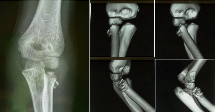
A 07 years old boy was admitted to emergency for left elbow trauma evolving for 24 hours. The patient felt at home when gaming on a table with landing on an outstretched hand. There was no particular surgical or medical history. We have at examination a painful elbow swelling with elective pain when touching the external proximal left forearm. Neurological examination was normal. X-ray showed a fracture with a bone fragment probably detached from the radius neck, without an elbow dislocation sign (Figure 1A). Computed tomography showed an epiphyseal-metaphyseal fracture line interesting the radial proximal extremity concluding therefore to Salter-Harris type 4 radial proximal injury. The fracture angulation was > 30° with a translation > 03mm (Figure 1B). Management procedure was explained to the parents but they did not give their consent for it and discharged.

Figure 1: (A) x-ray image showing a fracture with a bone fragment detached from the radial proximal extremity, (B) left elbow CT image showing an epiphyseal-metaphyseal fracture line (Salter-Harris type 4).
Discussion
Radial neck fractures are more prevalent at ages 09 to 10 years. At this age, the radial proximal physis is not close. Damage to this physis most often determines a Salter-Harris type 2 injury [1,3]. The Salter-Harris type 4 (epiphyso-metaphyseal) injury remains exceptional [3]
The mechanism of injury is usually a fall on an outstretched hand with a valgus compressive force across the elbow joint. There can also be a torsional component to the injury. Neurological examination is important as the posterior interosseous nerve is at risk due to its proximity to the radial neck. Complications of this fracture are elbow stiffness (most common) or partial physeal arrest which can affect the length and alignment of the radius in the future (cubitus valgus) [2].
Angulation and translation in radial neck fracture determine the type of treatment. If angulation is less than 30° and translation is less than 03 mm, closed reduction with immobilization in a long-arm cast is acceptable. Operative repair is usually indicated when the fracture angulation is greater than 30 degrees (following attempts at a closed reduction), translation of greater than 3 mm, and the reduced range of supination and pronation of under 45 degrees [2].
This operative repair of this injury (Salter-Harris type 4) means is:
- Closed percutaneous reduction using a K-wire or the Metaizeau technique using an elastic retrograde intramedullary nail [1,2].
- Open reduction by lateral skin incision (5-6 cm gently curved skin incision over the lateral condyle, elbow at 90 degrees or alternatively) or a posterolateral incision (behind the supracondylar ridge to the lateral border of the ulna) followed by a K-wire fixation or elastic stable intramedullary nailing (Metaizeau technique) [1,2].
Open reduction should only be used if closed procedure fails because open procedure, complications such as avascular necrosis, non-union, and radio-ulnar synostosis can occur [1,3].
Conclusion
Radial neck fracture is rare in children. A Salter-Harris type 4 injury remains exceptional at that location. The therapeutic choices in this injury are based on certain well-defined criteria.
Consent: written informed consent was obtained from the patient’s parents for publication of this case report and accompanying images. A copy of the written consent is available for review by the Editor-in-Chief of this journal on request.
Ethical approval: Ethical Approval was provided by the authors institution
Funding: no funding or grant support.
Guarantor: Sosso Piham KEBALO
Conflict of Interest statement: None
References
- Khajuria A, Tiwari V. Radial Neck Fracture Repair in A Child. In: StatPearls [Internet]. Treasure Island (FL): StatPearls Publishing, 2022.
- Pring ME. Pediatric Radial Neck Fractures. J Pediatr Orthop, 2012; 32 suppl (1): S14–S21. doi: 10.1097/BPO.0b013e31824b251d
- Lascombes P, Haumont Th. Radial neck fracture. In: fractures of the child. Montpellier: sauramps medical, 2002. 137-141.
- Agha RA, Franchi T, Sohrabi C, Mathew G, for the SCARE Group. The SCARE 2020 Guideline: Updating Consensus Surgical CAse REport (SCARE) Guidelines, International Journal of Surgery, 2020; 84: 226-230.

