Third Degree Uterovaginal Prolapse in Pregnancy
Oranye BC, Ogunmokun A, Obienu M, Nwabulue A and Edemi R
Department of Obstetrics and Gynecology, Eko Hospital, Nigeria
Received Date: 22/01/2022; Published Date: 08/02/2022
*Corresponding author: Benjamin C Oranye, Department of Obstetrics and Gynecology, Eko Hospital, Lagos, Nigeria
Abstract
Uterine Prolapse in pregnancy is not common and early diagnosis is crucial to preventing complication. Complications may occur in the antenatal, intrapartum and /or post-natal period and may include miscarriage, cervical infection, preterm contractions, premature rupture of membranes, preterm labor and even maternal mortality. Management options include use of vaginal pessary, careful conservative management of pregnancy. Delivery mode is usually individualized. Vaginal or caesarean delivery may be used.
Keywords: Delivery; Vaginal pessary; Cerclage; Pregnancy; Genital Prolpase
Introduction
Uterine prolapse is the descent of the uterus and cervix from their anatomical positions into the vagina or introitus. It is caused by weakened pelvic floor muscles and ligaments that are unable to support the uterus and its contents. Although it may occur at any age, it is more common among postmenopausal women, multiparous women, and women who have undergone instrumental deliveries. Other predisposing factors include macrosomic neonate delivery, chronic cough, constipation, pregnancy, previous pelvic surgery, and Hispanic or white mother [1]. Clinical features of uterovaginal prolapse are a sense of heaviness in the vagina, foul-smelling discharge, vaginal protrusion, urinary symptoms, dyspareunia, and difficulty in defecation.
Case Presentation
A 30-year-old pregnant mother (gravida 2, para 1) at 15 weeks gestational age, whose expected date of delivery was on 05/20/2019, presented to our gynecological outpatient clinic with complaints of recurrent protrusion of the uterine cervix into the vagina for 5 weeks and vaginal spotting for 2 days. The protrusion was first noticed after the last confinement roughly 15 months before this current episode.
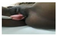
Initially, the vaginal swelling spontaneously decreased until 5 weeks before her clinical visit when it had increased in size, became irreducible and painful, and resulted in spotting. She had no history of prolonged labour, connective tissue disorder, macrosomic neonate delivery, or forceps use in her last delivery. She also had no recent urination difficulty or fever. No other significant complaints were noted. Ultrasound revealed a viable intrauterine pregnancy compatible with the gestational age. However, the entire cervix was outside the introitus. Nonetheless, no excoriations or ulceration was observed. The cervix was 2 cm dilated. Thus, she was diagnosed with third degree uterovaginal prolapse in pregnancy complicated with threatened miscarriage, thereby prompting admission. See Figures 1 and 2.


Discussion
Uterine prolapse occurs in 1 in 10,000-15,000 pregnancies; therefore, it is considered rare in pregnancy [2]. It can cause a feeling of heaviness in the pelvis and swelling at the introitus, causing difficulties in walking, sitting, and lifting objects [3], as in our patient. Complications may occur in the antepartum, intrapartum, or puerperal period and may include cervical infection, spontaneous miscarriage, urinary retention, urinary tract infection, preterm labor, cervical dystocia [4], and even maternal mortality [5,6]. Its mode of treatment and management may be varied. The delivery may be via vaginal delivery or cesarean section [7]. Vaginal pessary may also be inserted after prolapse reduction [8]. Our patient underwent prolapse reduction and vaginal ring pessary insertion after cervical cerclage placement. A successful pregnancy outcome requires individualized treatment with respect to patient’s wish, gestational age, and prolapse severity [9]. Continual use of a pessary is recommended until the onset of labor [10,11].
Conclusions
Uterine prolapse in pregnancy is a rare but challenging condition that may occur in gynecological outpatients. It presents with various symptoms and signs that could lead to pregnancy loss. Management includes cerclage use and vaginal pessary placement to support the pregnancy; close and serial ultrasound monitoring of the pessary, cerclage, and fetal growth; and symptom review. Elective cesarean section is recommended to prevent uterine prolapse during labor, as in the case of our patient.
References
- Uterine Prolapse: Symptoms and Causes, 2020.
- De Vita D, Giordano S. “Two: Successful Natural Pregnancies in a Patient with Severe Uterine Prolapse: A Case Report”. Journal of Medical Case Reports, 2011; 5: 459. DOI: 10.1186/1752-1947-5-459.
- Hardee K, Gay J, Blanc AK. Maternal morbidity: Neglected Dimension of Safe Motherhood in the Developing World, Global Public Health. 2012; 7: 603-617. DOI: 10.1080/17441692.2012.668919.
- Panagiotis T, Alexandros D, Nikolaos V, et al. Uterine Prolapse in Pregnancy: Risk Factors, Complications and Management. Journal of Maternal-Fetal & Neonatal Medicine, 2014; 27: 297-302. DOI: 10.3109/14767058.2013.807235.
- Andrea T, Antonio M, Siavash R, et al. Age-related Pelvic Floor Modifications and Prolapse Risk Factors in Postmenopausal Women. Menopause. 2010, 17:204-212. DOI: 10.1097/gme.0b013e3181b0c2ae.

