Multicystic Encephalomalacia
Sara Essetti*, Chaymae Faraj, Rachida Chehrastane, Sara Ez-zaky, Nazik Allali, Siham El Haddad and Latifa Chat
Pediatric Radiology Department, Children’s Hospital, Mohammed V University, Rabat, Morocco
Received Date: 11/06/2024; Published Date: 11/10/2024
*Corresponding author: Sara Essetti, Pediatric Radiology Department, Children’s Hospital, Mohammed V University, Rabat, Morocco
Clinical Image
A 5-month-old boy was born prematurely at 34 weeks gestation and was followed for hypoxic ischemic encephalopathy. During follow-up, delayed development was noted and MRI was performed for further evaluation.
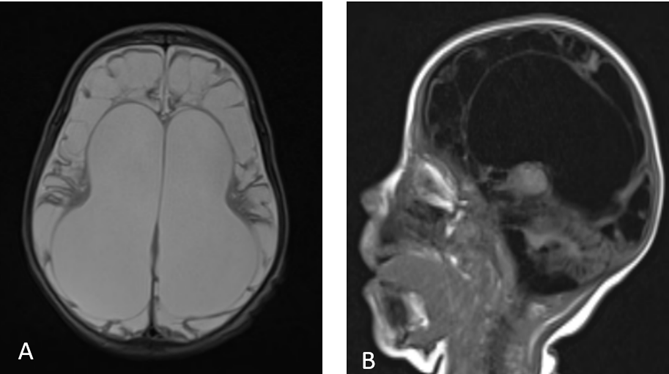
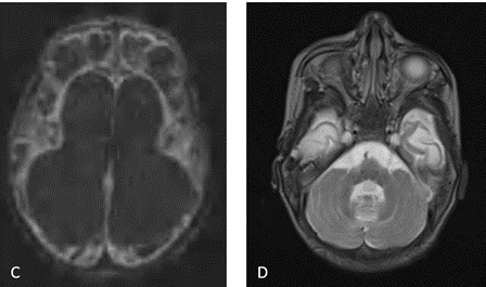
Brain MRI (Figure 1) showed multiple well-defined cystic cavities of varying size and shape in both cerebral hemispheres, involving both gray and white matter but sparing the brainstem and cerebellum; these cavities had low signal on T1, high signal on T2, and no restricted diffusion. Dilatation of the ventricular and atrophy of the corpus callosum were also noted. The diagnosis of multicystic encephalomalacia was made.


Figure 1: Brain MRI in axial and sagittal planes; T2WI (A), T1WI (B), and DWI (C): Multiple cystic areas of varying size in the cortex and white matter of both hemispheres with dilatation of the ventricular system and atrophy of the corpus callosum. Note the preserved posterior fossa (D).
Discussion
Multicystic encephalomalacia is a condition in which there are necrotic areas that develop into cystic lesions in the brain. It may be located in the cortical or white matter regions. It is commonly seen in term neonates as a result of extensive brain insult from asphyxia due to hypoxic-ischemic injury [1]. Other causes such as meningoencephalitis [2], twin-to-twin transfusion and abusive head trauma [3] may also result in this condition.
The location of the lesions depends on the nature of the insult [4]. If caused by thromboembolic infarction, the affected area will be in the distribution of a major cerebral artery. If the insult is due to partial asphyxia, the lesions are more likely to be in the cortex and peripheral white matter, especially in the watershed areas. If the insult is very severe, only the immediate periventricular white matter may be spared. There is usually marked ventriculomegaly because of ex vacuo dilatation from the extensive surrounding white matter destruction.
Transfontanellar ultrasound is the first imaging modality. It showed cortical atrophy with enlarged ventricles, cortical hyperechogenicity, and multiple cystic formations within the ventricles. On CT scan, cystic encephalomalacia is seen as a region of hypoattenuation with cysts of various sizes with volume loss. On MRI, it follows the CSF signal on all sequences, unlike gliosis, which appears bright on both T2 and FLAIR.
Multicystic encephalomalacia needs to be differentiated from hydranencephaly and porencephaly, by the presence of multiple cysts involving multiple lobes separated by glial septa [4].
Multicystic encephalomalacia is associated with a profound neuromotor retardation and, consequently, a serious prognosis.
References
- Bano S, Chaudhary V, Garga UC. Neonatal Hypoxicischemic Encephalopathy: a radiological review. J Pediatr Neurosci, 2017; 12(1): 1-6. http://dx.doi. org/10.4103/1817-1745.205646.
- Lane LM, McDermott MB, O’Connor P, et al. Multicystic Encephalomalacia: The Neuropathology of Systemic Neonatal Parechovirus Infection. Pediatric and DevelopmentalPathology, 2021; 24(5): 460-466. doi:10.1177/10935266211001645.
- Matlung SE, Bilo RA, Kubat B, van Rijn RR. Multicystic encephalomalacia as an end-stage finding in abusive head trauma. Forensic Sci Med Pathol, 2011; 7(4): 355-363. http://dx.doi.org/10.1007/s12024-011-92367.
- Folkerth RD. The neuropathology of acquired pre- and perinatal brain injuries.SeminDiagnPathol, 2007; 24(1): 4857. http://dx.doi.org/10.1053/j.semdp.2007.02.006.

