Cutaneous Presentation of Metastatic Acinic Cell Carcinoma
Hafsa El Boukili*, Sara Elloudi, Zakia Douhi, Meriem Soughi, Hanane Baybay, Fatima Zahra Mernissi
Department of Dermatology, University Hospital Hassan II, Faculty of Medicine and Pharmacy, Sidi Mohamed Ben Abdellah University, Morocco
Received Date: 23/03/2024; Published Date: 19/08/2024
*Corresponding author: Hafsa El Boukili, Department of dermatology, University Hospital Hassan II, Faculty of Medicine and Pharmacy, Sidi Mohamed Ben Abdellah University, Fez, Morocco
Description
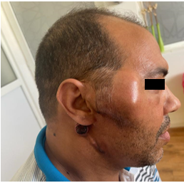
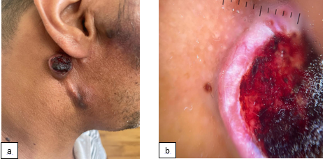
A 39-year-old man, who had been under observation for Acinic Cell Carcinoma of the parotid gland 4 years prior, he underwent a right parotidectomy. Initial oncologic management conducted multiple treatment cycles with several chemotherapeutic agents over the course of 03 years. Presented with a rapidly enlarging, tender and asymptomatic retro auricular nodule. Clinical examination revealed a smooth mass measuring 70 mm×60 mm in preauricular right region, it was, firm without signs of tenderness, and an erythematous slightly elevated nodule on the right mastoid area, sharply demarcated, with a surface covered by hemorrhagic crusts, and crater-like appearance (Figure 1) On dermoscopy: Peripheral arborescent vessels; white structureless area were seen (Figure 2). Given the dubious appearance, whether it is a keratoacanthoma or a cutaneous metastasis, a punch biopsy was indicated, and the diagnosis of acinic cell carcinoma metastatic to the skin was established. Due to the tumor progression, the patient was considered eligible for radiotherapy.

Figure 1: Face of patient with right parotid gland tumor and mastoid nodule.

Figure 2: (a) Clinical aspect of the nodule (b) Dermoscopic aspect showed Peripheral arborescent vessels; white structureless area.
Teaching Point
The documented occurrence of cutaneous metastases originating from a confirmed primary malignancy varies from 0.6% to 9% [1]. In spite of being a low‑grade malignant tumor, Acinic Cell Carcinoma (ACC) has a propensity for late recurrence and metastasis, often many years after initial presentation. Most common sites of metastasis are the lungs, brain, and lymph nodes, Case reports of isolated concurrent cutaneous involvement have been reported [2]. The possibility of metastatic ACC should be considered in the differential diagnosis of new cutaneous nodules in patients with a history of ACC. The presence of detectable vascular structures within a skin nodule in individuals already diagnosed with cancer should prompt concern for cutaneous metastasis. The frequent occurrence of vascular structures in cutaneous metastases implies a potential involvement of angiogenesis in their development [3] these results endorse the utilization of dermoscopy to assess suspected skin metastases.
References
- Lookingbill DP, Spangler N, Helm KF. Cutaneous metastases in patients with metastatic carcinoma: a retrospective study of 4020 patients. J Am Acad Dermatol, 1993; 29(2, pt 1): 228-236.
- Ashini Shah, Shailee Mehta, Amisha Gami, Dhaval Jetly. Acinic Cell Carcinoma of Parotid Gland Presenting as Disseminated Cutaneous and Subcutaneous Metastasis after 20 Years of Initial Presentation. Indian Journal of Medical and Paediatric Oncology, 2019; 4 (Supplement 1).
- Chernoff KA, Marghoob AA, Lacouture ME, Deng L, Busam KJ, Myskowski PL. Dermoscopic Findings in Cutaneous Metastases. JAMA Dermatology, 2014; 150(4): 429.

