Clinical and Dermoscopic Features of Breast Sebaceous Adenoma
Hafsa El Boukili*, Hanane Baybay, Zakia Douhi, Meryem Soughi, Sara Elloudi and Fatima Zahra Mernissi
Department of Dermatology, University Hospital Hassan II, Faculty of Medicine and Pharmacy, Sidi Mohamed Ben Abdellah University, Morocco
Received Date: 23/03/2024; Published Date: 16/08/2024
*Corresponding author: Hafsa El Boukili, Department of dermatology, University Hospital Hassan II, Faculty of Medicine and Pharmacy, Sidi Mohamed Ben Abdellah University, Fez, Morocco
Presentation
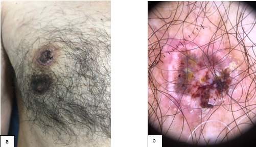
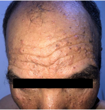
A 79-year-old, man presented a painless lesion with progressive growth on the right breast. His past medical history included: a squamous cell carcinoma of the lower lip 10 months prior, he underwent an excision surgery with reconstruction. On Examination: a red-yellowish tumor of about 2 cm, with multiple telangiectasias, irregular surface, erosion, and crust was observed on his right nipple (Figure 1a), dermoscopy showed multiple pale yellow globules on an erythematous background, besides tortuous ramified vessels, more evident in the periphery and central crust (Figure 1b), we also noted multiple pink-white papules on the face (Figure 2) suggesting a pattern of sebaceous gland neoplasia, irritated seborrheic keratosis and trichelemmal carcinoma were also considered as relevant differential diagnoses. Subsequently, the lesion was excised, and histopathological examination confirmed the diagnosis of sebaceous adenoma. At the time of diagnosis, there were no indications of associated malignancy.

Figure 1: (a) Red-yellowish irregular tumor of about 2 cm, observed up on the right nipple (b) Pale yellow globules on an erythematous background, besides tortuous ramified vessels, more evident in the periphery.

Figure 2: Multiple pink-white papules on forehead and cheeks.
Teaching Point
Sebaceous adenoma is a multilobular tumor with sebaceous differentiation, it can present as an isolated or multiple lesions, either dome-shaped, papule or nodule, rarely ulcerated, ranging between 0.5 and 10 cm in diameter, with yellowish color, located mainly on the face or scalp [1]. When associated with gastrointestinal or genitourinary neoplasms, the sebaceous adenoma can be part of Muir–Torre syndrome. It is advised that these patients should submit to a more detailed investigation both at the time of the diagnosis and during periodic follow-up. Many dermatoscopic criteria for sebaceous hyperplasia have already been well documented, distinguishing tumors with a central crater, characterized by crown vessels that embrace an opaque structureless ovoid white-yellow center, an aspect wich is similar to the present case, and tumors without a central crater, which reveal dermoscopically branching but unfocussed arborizing vessels over a white to yellow background and few loosely arranged yellow comedo-like globules [2]. Hence, the examination of a larger number of lesions is likely to establish more specific criteria for diagnosis in the future.
References
- Moscarella E, Argenziano G, Longo C, et al. Clinical, dermoscopic and reflectance confocal microscopy features of sebaceous neoplasms in Muir-Torre syndrome. J Eur Acad Dermatol Venereol, 2013; 27(6): 699-705. doi: 10.1111/j.1468-3083.2012.04539.x.
- Marques-da-Costa J, Campos-do-Carmo G, Ormiga P, Ishida C, Cuzzi T, Ramos-e-Silva M. Sebaceous adenoma: clinics, dermatoscopy, and histopathology. International Journal of Dermatology, 2014; 54(6): e200–e202. doi: 10.1111/ijd.12722.

