Demoscopy Sheds Light on Perioral Lesion Mimcking Herpes
Ghita Sqalli Houssini*, Hanane Baybay , Zakia Douhi, Meryem Soughi, Sara Elloudi and Fatima Zahra Mernissi
Department of Dermatology, University Hospital Hassan II, Faculty of Medicine and Pharmacy, Sidi Mohamed Ben Abdellah University, Morocco
Received Date: 13/03/2024; Published Date: 01/08/2024
*Corresponding author: Dr. Sqalli Houssini Ghita, Department of Dermatology, University Hospital Hassan II, Faculty of Medicine and Pharmacy, Sidi Mohamed Ben Abdellah University, Fez, Morocco
Observation
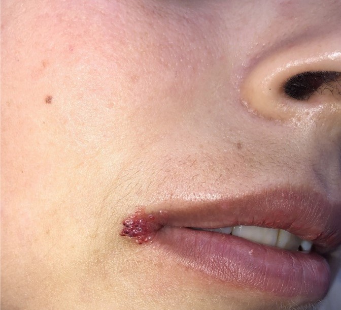
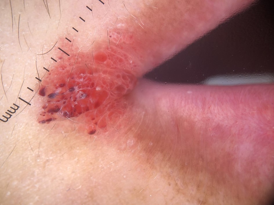
A 31-year-old woman with a niece who was treated for a venous malformation consulted us for management of a perioral lesion that had been present since childhood. She had been treated with valacyclovir as an herpes on several occasions without improvement. Our examination revealed vesicles grouped in clusters with hemorrhagic content in the perioral area. Dermoscopy revealed red lacunae and a hypopyon aspect, leading to the diagnosis of perioral microcystic lymphangioma. We decided to treat her with a vascular laser.

Figure 1: Presence of red vesicles clustered in a bouquet at the peribuccal level.

Figure 2: Dermoscopy of the peribuccal lesion reveals red lacunae with the hypopyon sign.
Teaching Point
Diagnosing lymphangioma circumscriptum is usually straightforward based on its clinical presentation and behavior. While the majority of cases exhibit typical features, there may be instances of solitary lesions or atypical appearances. Dermoscopy plays a crucial role in ensuring an accurate diagnosis, with common dermoscopic features including lacunae, vascular structures, white lines, and the hypopyon sign [1-3]. Lacunae manifest as multiple, clustered, well-defined, and variably colored structures with a round to oval shape [1]. Additionally, the hypopyon sign may be observed, characterized by a two-tone lacuna or a color transition from dark (at the bottom) to light (at the upper part) within the same lacuna. This phenomenon is attributed to blood sedimentation in the dilated lymphatic channels [1-3]. The presence of the hypopyon sign and lacunae strongly indicates lymphangioma circumscriptum, underscoring the significance of a comprehensive dermoscopic examination in cases of chronic lesions that resist typical treatments. It is crucial not to misinterpret any perioral lesion with vesicles as a simple case of herpes
Consent : The examination of the patient was conducted according to the Declaration of Helsinki principles.
Conflict of Interest: The authors do not declare any conflict of interest.
References
- Zaballos P, Del Pozo LJ, Argenziano G, Karaarslan IK, Landi C, Vera A, et al. Dermoscopy of Lymphangioma Circumscriptum: A morphological study of 45 cases. Australas J Dermatol, 2018; 59(3): e189-e193.
- Kabbur G, Jaimes JP. Hyphema-like sign in dermatoscopy of a lymphangioma. JAAD Case Rep, 2020; 6(10): 959-960.
- Massa AF, Menezes N, Baptista A, Moreira AI, Ferreira EO. Cutaneous Lymphangioma circumscriptum - dermoscopic features. An Bras Dermatol, 2015; 90(2): 262-264.

