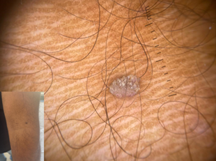
Asymptomatic Dark Grey Papule: What Dermoscopy had Revealed
El Boukili Hafsa*, Douhi Zakia, Soughi Meryem, Elloudi Sara; Baybay Hanane; Mernissi Fatima Zahra
Department of Dermatology and Venerology, University Hospital Hassan II, Morocco
Received Date: 24/01/2024; Published Date: 06/06/2024
*Corresponding author: El Boukili Hafsa, Department of Dermatology and Venerology, University Hospital Hassan II, Fez, Morocco
Case Presentation
A 32-year-old male consulted for a painless lesion on the right knee, which had been evolving for 3 months; initially, he denied any application of a topical product. Upon examination, a blackish-grey papule, 15 mm in diameter, with a rough surface and pigmented border was noted. Faced with the suspicious blackish appearance, we used the dermatoscope on it: Hemorrhagic points and lines surrounded by a whitish halo were seen, this combination of features gives an appearance reminiscent of frogspawn (Figure 1), the rest of the dermatological examination revealed on the left arm a cluster of yellowish fine fronds emerging from a narrow pedicle base (Figure 2). Detailed questioning with the patient revealed the notion of the application of silver nitrate into the first lesion, which could explain the black appearance observed. Considering these findings, the diagnosis of warts was made.

Figure 1: Hemorrhagic points and lines surrounded by a whitish halo, known as “Frogspawn pattern”.

Figure 2: Finger like projections with bleeding spots on it.
Teaching point
Cutaneous warts caused by Human papilloma virus can affect up to 7–12% of the general population [1]. They are benign proliferations of the epidermis and may present in different forms; The diagnosis of this viral infection is usually made clinically, but dermoscopy can be of aid when the diagnosis is uncertain. The two aspects mentioned above have been described in the literature [2] as a “frogspawn” pattern, which shows multiple polyps resembling a mass of frog eggs in the first lesion; and the Finger like projections in the filiform wart observed in the second lesion. Therefore, the use of dermoscop was sufficient to confirm the diagnosis of a wart and reassure the patient about the blackish appearance of the first lesion, which is due to the exogenous deposition of silver nitrate.
References
- Clifton MM, Johnson SM, Roberson PK, Kincannon J, Horn TD. Immunotherapy for recalcitrant warts in children using intralesional mumps or candida antigens. Pediatr Dermatol, 2003; 20: 268–271.
- Zalaudek I, Giacomel J, Cabo H, Di Stefani A, Ferrara G, Hofmann-Wellenhof R, et al. Entodermoscopy: A New Tool for Diagnosing Skin Infections and Infestations. Dermatology, 2007; 216(1) : 14–23. doi:10.1159/000109353.

