Corpus Callosum Agenesis: The Typical Racing Car Sign
Essetti S*, Faraj C, El Harras Y, Chait F, Allali N, El Haddad S and Chat L
Department of Pediatric and Gynecology Radiology, Children's hospital, University Mohammed V, Rabat, Morocco
Received Date: 08/06/2023; Published Date: 20/10/2023
*Corresponding author: Essetti Sara, Department of Pediatric and Gynecology Radiology, Children's hospital, University Mohammed V, Rabat, Morocco
Corpus callosum is a white matter tract that connects the cerebral hemispheres, facilitating interhemispheric connectivity. It has four parts: the rostrum, the genu, the body, and the splenium. Its development starts in the first trimester of pregnancy, while maturation continues in childhood and adolescence [1].
Anomalies in the development of corpus callosum vary from partial (dysgenesis) to complete (agenesis). MRI is the modality of choice for diagnosis [2].
Clinically, partial dysgenesis can be asymptomatic, while children with agenesis usually have developmental defects, seizures, psychomotor or mental retardation.
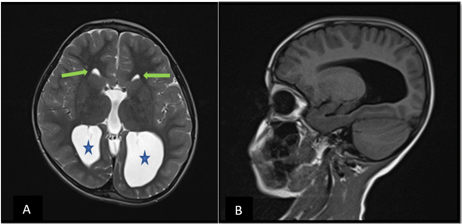
The “racing car” sign is an MRI sign which can be seen in complete agenesis of corpus callosum, widely spaced anterior horns of the lateral ventricles representing the anterior wheeling tires, and the dilated posterior horns representing the posterior driving tires (Figure 1: A).
It is primordial for radiologists to be aware of the features of this pathology in order to distinguish it from hydrocephalus. Furthermore, to instigate a search for other, more serious, anomalies with which it may be associated, since it can highly vary the overall prognosis. Treatment usually includes conservative management with rehabilitation.

Figure 1: Cerebral MRI of a 5 years old girl
A: Axial T2 weighted image showing widely near-parallel spaced frontal horns of the lateral ventricles (green arrow) and teardrop-shaped dilatation of their posterior trigones (colpocephaly ) reminiscent of a racing car.
B: Sagittal T1 weighted image showing a complete absence of corpus callosum.
References
- Paul LK, Brown WS, Adolphs R, Tyszka JM, Richards LJ, et al. Agenesis of the corpus callosum: genetic, developmental and functional aspects of connectivity. Nat Rev Neurosci, 2007; 8: 287-299.
- Manfredi R, Tognolini A, Bruno C, Raffaelli R, Franchi M, et al. Agenesis of the corpus callosum in fetuses with mild ventriculomegaly: role of MR imaging. Radiol Med, 2010; 115: 301-312.

