Splenic Artery Aneurysm Incidentally Discovered on Echo-Endoscopy
Alae Chakir1,*, Tonguino RN1, Elkouti I1, Yousfi H1, Belabas S2, Bentaher A2 and Lamsiah T1
1Department of Gastroenterology, Moulay Ismail Military Hospital- Meknes, Morocco
2Department of Radiology, Moulay Ismail Military Hospital- Meknes, Morocco
Received Date: 30/12/2022; Published Date: 25/01/2023
*Corresponding author: Alae Chakir, Department of Gastroenterology, Moulay Ismail Military Hospital- Meknes, Morocco
Abstract
Splenic artery aneurysm is a rare entity, Most often asymptomatic, we report the case of a patient with fortuitous discovery of a splenic artery aneurysm on an echo-endoscopy image confirmed by angiography scan.
Keywords : Splenic artery aneurysm; Angiography scan; Echo-endoscopy; Thrombosis
We report the case of a 56-year-old patient, cholecystectomized 3 months ago, followed for Churg Strauss vasculitis under oral corticosteroid therapy, admitted for intermittent hepatic colic with disturbances of the hepatic assessment: cytolysis with slight cholestasis.
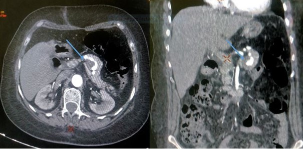
An echo-endoscopy was carried out as part of the search for a persistent calculation in the main bile duct revealed the presence of a 3 cm aneurysm of the splenic artery with partial thrombosis (Figure 1). An abdominal angiography scan with three-dimensional reconstructions was requested for a better characterization, which showed the image of a partially thrombotic splenic artery aneurysm (Figure 2 and 3).
Splenic artery aneurysm is a rare entity with an incidence of 0.01-0.2% [1]. Most often asymptomatic and their discovery is made fortuitously ,during the realization of an abdominal morphological examination indicated for another pathology [2] .The angiography scan makes it precise lesional assessment by determining the dimensions of the aneurysm, and by distinguishing between the true lumen and the parietal thrombus [3].
The digestive echo-endoscopy is a reference examination for the exploration of the biliopcreatic crossroads, the combined use of the Doppler also makes it possible to visualize the abdominal arteries and veins, and this is the case of our patient whose examination allowed us to detect this aneurysm of the splenic artery.

Figure 1: Echoendoscopic image of a partially thrombosed splenic artery aneurysm with Doppler showing arterial flow.

Figure 2: Scannographic sections showing a partially thrombosed splenic artery aneurysm.

Figure 3: Three-dimensional reconstruction images showing a splenic artery aneurysm.
References
- Reardon PR, Otah E, Craig ES, Matthews BD, Reardon MJ. Résection laparoscopique des anévrismes de l'artère splénique. Chirurgie Endosc. Avr, 2005; 19(4): 488–493.
- Abbas MA, Stone WM, Fowl RJ, Gloviczki P, Oldenburg WA, Pairolero PC, et al. Anévrismes de l'artère splénique : deux décennies d'expérience à la clinique Mayo. Ann Vasc Surg. Juill, 2002; 16(4): 442–449.
- Wolinski AP, Gall WJ, Dubbins PA. Anévrysme de l'artère hépatique suite à une pancréatite diagnostiquée par échographie. Revue britannique de radiologie, 1985; 58(692): 768–770.

