Airway Obstruction as the First Manifestation of an Anaplastic Thyroid Carcinoma
Fábia Cruz1,*, Rita Branquinho Pinheiro2, Rita Macedo3, João Dias Cardoso4, Francisco Freitas4, Paula Monteiro5, Paula Pinto6 and Cristina Bárbara7
1Internal Training Inpatient, Integrated Responsibility Center for Internal Medicine, Hospital Amato Lusitano, ULSCB, Portugal
2Internal Specific Training in Pulmonology, Hospital Santa Maria, HCF/CHULN, Portugal
3Pulmonology Hospital Assistant, Hospital Santa Maria, CHULN, Portugal
4Pulmonology Hospital Assistant, Intervention Pulmonology Unit, Hospital Santa Maria, CHULN, Portugal
5Graduate Hospital Assistant of Pulmonology, Intervention Pulmonology Unit, Hospital Santa Maria, CHULN, Portugal
6Graduate Hospital Assistant of Pulmonology, Hospital Santa Maria, CHULN. Faculty of Medicine of Lisbon, ISAMB, Portugal
7Senior Graduated Hospital Assistant of Pulmonology, Hospital Santa Maria, CHULN. Faculty of Medicine of Lisbon, ISAMB, Portugal
Received Date: 15/09/2022; Published Date: 10/10/2022
*Corresponding author: Fábia Cruz, 1Internal Training Inpatient, Integrated Responsibility Center for Internal Medicine, Hospital Amato Lusitano, ULSCB, Portugal
Case Description
Anaplastic Thyroid Carcinoma (ACT) is a rare malignancy, accounting for 1-2% of all thyroid cancers. Although rare, ATC accounts for most of deaths from thyroid carcinoma.
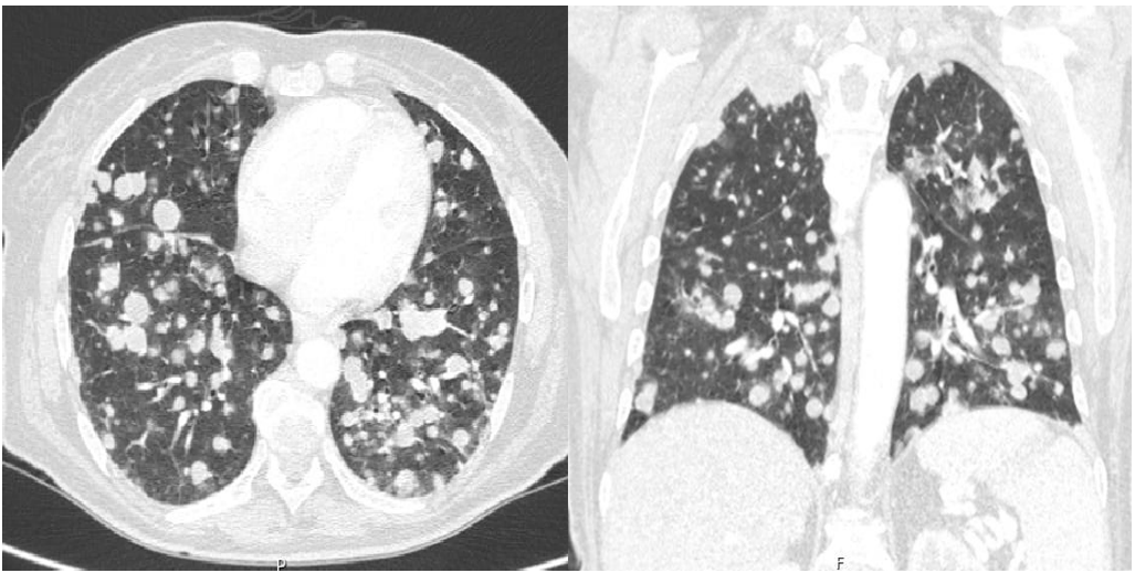
A 64-years-old woman present in the emergency department due to solid dysphagia, “throat tightness”, hoarseness and hemoptoic sputum with one week. A mass was identified at the base of the neck (Figure 1), adherent to the planes and painless. Thyroid ultrasound revealed large solid hypoechoic lesion in the right lobe extending to the isthmus, with irregular contours, classified as EU-TIRADS 5 (European Thyroid Association Guidelines for Ultrasound Malignancy Risk Stratification of Thyroid Nodules in Adults). Chest CT scan was suggestive of bilateral metastases (Figure 2). Rigid bronchoscopy was performed to removal intratracheal mass with immediate symptomatic relief (Figure 3). Pathological anatomy revealed anaplastic thyroid carcinoma.
Anaplastic carcinoma is fast-growing, and early diagnosis is essential to improve its reserved prognosis.
Keywords: Anaplastic thyroid carcinoma; Upper airway obstruction

Figure 1: Mass at the base of the neck.

Figure 2: Chest CT scan with bilateral metastases.

Figure 3: Rigid bronchoscopy show a intratracheal mass, 1.5cm below vocal cords.
Patient's consent: Written informed consent was obtained for the publication in this case report and accompanying images.
Funding: The authors declare that no funding was received for this article.
Declaration of Competing Interest: None of the authors have any conflicts of interest to disclosure.
Authors Contribution
Fábia Cruz: Acquisition of data, drafting the article and literature revision Rita Branquinho Pinheiro: acquisition of data
Rita Macedo: Literature revision João Dias Cardoso: critical revision Francisco Freitas: critical revision Paula Monteiro: guarantor
Paula Pinto: Guarantor
Cristina Bárbara: Guarantor
References
- Molinaro E, Romei C, Biagini A, Sabini E, Agate L, Mazzeo S, et al. Anaplastic thyroid carcinoma: from clinicopathology to genetics and advanced therapies. Nat Rev Endocrinol, 2017; 13(11): 644-660.
- Tiedje V, Stuschke M, Weber F, Dralle H, Moss L, Führer D. Anaplastic thyroid carcinoma: review of treatment protocols. Endocr Relat Cancer, 2018; 25(3): R153-R161.
- Manzoor D, Balzer BL, Gayhart M, Vail E, Marchevsky AM, Setoodeh R. Anaplastic thyroid carcinoma presenting as laryngotracheal invasive squamous cell carcinoma: A report of two cases and review of the literature. Human Pathology: Case Reports, 2021; 24: 200505.
- Deeken-Draisey A, Yang GY, Gao J, Alexiev BA. Anaplastic thyroid carcinoma: an epidemiologic, histologic, immunohistochemical, and molecular single-institution study. Hum Pathol, 2018; 82: 140-148.
- Johnson J, Lai S, Cotzia P, Cognetti D, Luhinbuhl A, Pribitkin E, et al. Mitochondrial Metabolism as a Treatment Target in Anaplastic Thyroid Cancer. Seminars in Oncology, 2015; 42(6): pp 915-922.

