Hourglass Appearance Typical of Classic Lissencephaly
Choayb S*, Adil H, Allali N, Chat L and El Haddad S
Children’s Hospital, Radiology Department, Mohamed V University, Rabat, Morocco
Received Date: 09/06/2021; Published Date: 25/06/2021
*Corresponding author: Choayb Safaa, Children’s Hospital, Radiology Department, Mohamed V University, Rabat, Morocco 2020. Email: choayb.safaa@gmail.com
Keyword: lissencephaly – Hourglass appearance – child seizure
Observation
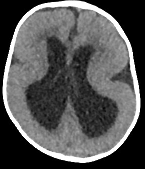
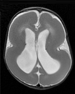
A 13-month-old girl presenting seizures and developmental delay. An initial head CT was performed showing a smooth cortical surface with no normal gyration (Figure 1). A brain MRI was preformed that shows the classic hourglass configuration of the cerebral hemispheres with the absence of cerebral sulcation (Figure 2).

Figure 1: Axial slice of non-contrast CT head shows an underformed gyri with a smooth cortical surface

Figure 2: Axial T2WI MR shows the typical hourglass or figure of eight appearance.
Comment
Lissencephaly is caused by arrested neuronal migration, resulting in a smooth brain surface, Type 1 or “classical” lissencephaly, is defined by a lack of gyri and sulci in the cerebral neocortex. Besides, there is a simplification of neocortical lamination. Typically, the neocortex of lissencephalic patients has three to four layers instead of six. [1]
It is caused by a variety of gene alterations; LIS1 and DCX are responsible for classic lissencephaly.
The disruption of neuronal migration occurs between the 10th and 14th weeks of gestation. Imaging findings range from the classic hourglass or figure of 8 shape due to shallow, vertically oriented Sylvian fissures, reflecting diffusely thickened and abnormally smooth cerebral cortex in its more severe form, to milder forms of subtle band heterotopias. More severe forms are manifested in infancy with hypotonia, seizures, and developmental delay. The less severe forms with only subcortical band heterotopia are more frequently observed in heterozygous females with an X-linked DCX gene mutation and may be less symptomatic with mild seizures and present later in childhood or in the adult setting. [2]
When suspecting lissencephaly in neonate, verify gestational age, Agyric (smooth) cortex is normal up to 26 weeks (MRI, US)
Declaration of Interests
The authors declare that they have no competing interest.
References
- Loturco J.J and Manent J. Neuronal migration disorders. Elsevier Inc., 2020.
- Choi J.J, Setty B.N, and Mph E.Y.L. B r a i n Ma l f o r m a t i o n s a t A l l Ages From Aunt Minnie to Zebras for General Radiologists. Radiol. Clin. NA, 2020.

