Osteolytic lesions in Multiple Myeloma
Marina Costa, Joana Medeiros, Rosário Araújo
Internal Medicine Department, Hospital de Braga, Portugal
Received Date: 17/02/2021; Published Date: 09/03/2021
*Corresponding author: Internal Medicine Department, Hospital de Braga, Braga, Portugal.
Case Description
An 83-year-old woman, with history of hypertension, dyslipidaemia and osteoarticular disease, was admitted to the internal medicine department with a 2-week history of dyspnoea, anorexia, dorsal pain and prostration. She presented with an acute kidney injury (plasma creatinine, 2.0 [normal 0.6-1.2] mg/dL), a metabolic alkalaemia (pH, 7.568 [normal 7.35-7.45]; HCO3-, 34.7 [normal 21-26] mmol/L) and a hypoxemic respiratory failure.
The following workup revealed the ensuing results: hemoglobin, 7.8 (normal 11.9-15.6) g/dL; platelet count, 82 (normal 150-140) x 103/uL; plasma calcium, 12.6 (normal 8.3-10-6) mg/dL; serum albumin, 2.4 (normal 3.4-5.0) g/dL; serum IgG, 4641 (normal 650-1600) mg/dL; free light Lambda chains, 395.00 (0.83-2.70) mg/dL and K/L ratio, 0.002 (normal 0.31-1.56); Beta2-microglobulin, 22687 (normal 1000-2400) ng/mL. The serum protein electrophoresis showed a spike on beta-2 region, 4.8 (normal 0.2-0.5) g/dL, representing 56.5% of serum proteins; and a monoclonal gammopathy IgG/Lambda demonstrated by immunoelectrophoretic.

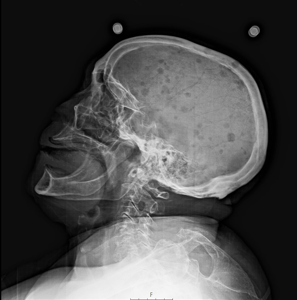
Figure 1: Radiograph of the skull showing multiple “punched out” radiolucent lesions in multiple myeloma.

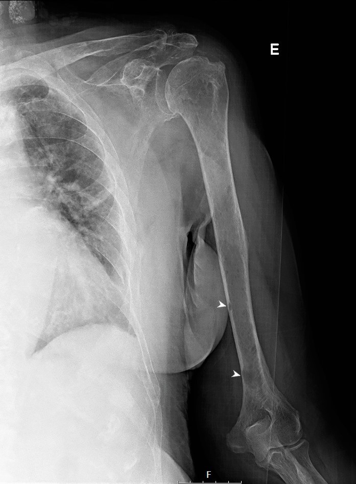
Figure 2: Radiograph of the humerus, with radiolucent well-defined lesions (white arrow heads) in multiple myeloma.

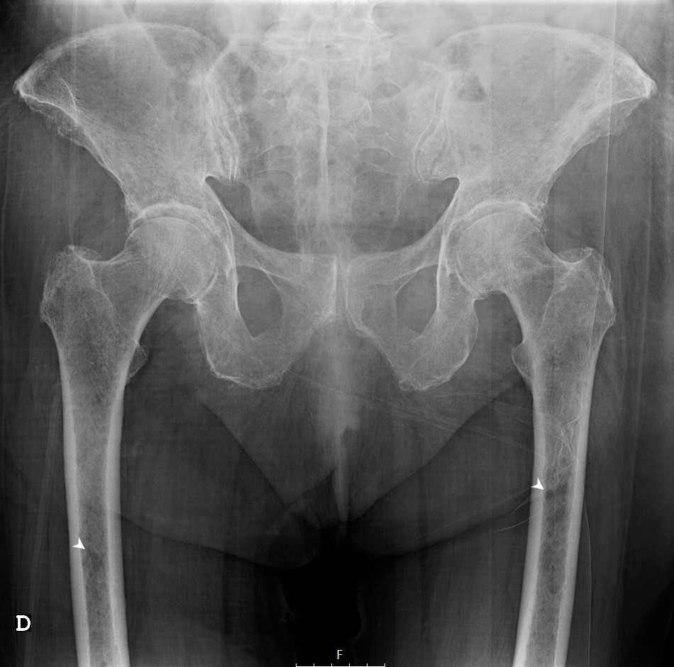
Figure 3: Radiograph of the hip with punched out lesions in both femur bones (white arrow heads).
Pursuing the hypothesis of Multiple Myeloma (MM), a whole-body skeletal X-ray was performed, showing characteristic multiple “punched out” radiolucent lesions on the skull (Figure 1), humerus (Figure 2) and pelvis (Figure 3), as a result of destruction by nodules of plasma cells. Bone marrow biopsy showed a diffuse interstitial infiltration by neoplastic cells from the plasmocytic lineage, comprising about 70% of the cell population, confirming the diagnosis of MM. The patient was reffered to a hemathologist but, despite adequate treatment, she deceased 4 months after diagnosis.
MM is one of the hardest cancers to diagnose, partially due to its non-specific presenting features [1]. Back pain is the most frequent symptom at presentation however, it is also the second most common complaint in the primary care setting, most of it not cancer-related, contributing to the delay in diagnosis of MM [2-4]. The late diagnosis is associated with a higher incidence of complications and worse disease-free survival [5].
Conflicts of Interest
The authors have no conflicts of interest to declare.
Grant Information
The authors received no specific funding for this work.
References
- Lyratzopoulos G, et al. Rethinking diagnostic delay in cancer: how difficult is the diagnosis? BMJ. 2014; 349: g7400.
- Goldschmidt N, et al. Presenting Signs of Multiple Myeloma and the Effect of Diagnostic Delay on the Prognosis. The Journal of the American Board of Family Medicine. November 2016; 29 (6): 702-709.
- Deyo RA, et al. What can the history and physical examination tell us about low back pain? JAMA. 1992; 268: 760–765.
- Jarvik JG, Deyo RA. Diagnostic evaluation of low back pain with emphasis on imaging. Ann Intern Med. 2002; 137: 586–597.
- Kariyawasan CC, et al. Multiple myeloma: causes and consequences of delay in diagnosis. QJM. 2007; 100(10): 635-640.

