Pulmonary Emphysema in Covid-19 Pneumonia: Ct Findings
Francesco MESSINA**, Annamaria NUCERA*, Caterina TRIPODI* and Nicola ARCADI*
*Unit of Radiology – Riuniti Hospital
Azienda Ospedaliera Grande Ospedale Metropolitano (G.O.M.) “Bianchi-Melacrino-Morelli” Reggio Calabria, Italy
Received Date: 04/06/2021; Published Date: 21/06/2021
*Corresponding author: Francesco Messina, MD, Unit of Radiology - Riuniti Hospital, Azienda Ospedaliera GrandeOspedale Metropolitano “Bianchi–Melacrino–Morelli” Via Giuseppe Melacrino n.21, 89124 Reggio Calabria, Italy.Email: fmessina1@hotmail.it, Phone number: +393392641419
Abstract
Since the widespread of acute respiratory syndrome infection caused by Coronavirus-19, chest computed tomography (CT) was considered a useful imaging tool commonly used in early diagnosis and monitoring of patients with Covid-19 pneumonia. Many typical imaging features of this disease were carefully described with chest CT, as well as the collateral CT findings in the lungs and mediastinum. In our case we describe a patient with bilateral Covid-19 pneumonia, that collaterally had a large and voluminous bulla of pulmonary emphysema in the left lung, documented at CT.
Keywords: Pulmonary emphysema Covid-19; Pneumonia; Computed Tomography
Background
Since December 2019 the world is facing a rapidly expanding pandemic of lower respiratory tract infection by a novel coronavirus SARS-CoV-2 (severe respiratory syndrome coronavirus-2). In some patients, this viral infection causes a clinical syndrome referred to as coronavirus disease 2019 (Covid-19), but the heterogeneity of the disease course poses a challenge to healthcare providers and optimal management of patients. Since most Covid-19 infected patients were diagnosed with pneumonia, chest CT played a central role in the diagnosis and management [1]. Several case series and case reports demonstrated the CT features on presentation and its temporal progressions during therapy. The use of CT imaging in the diagnosis and follow-up had rapidly grown, and radiological patterns along the disease course are increasingly understood. Chest CT imaging has been demonstrated more sensitive than chest radiography to identify the manifestations of Covid-19 pneumonia, for the severity assessment and monitoring of the disease [2,3].
Case Presentation
A 74 years old man presented at emergency department with a history of recent chest pain since about a week, with dry cough, shortness of breath and fever (37.5°C). He is diabetic and also suffers from pulmonary emphysema. He had been treated at home with paracetamol, without any improvement in his symptoms. Chest clinical auscultation revealed a bi-basal reduced intensity of breath sounds (more on the left). Arterial oxygen saturation (SaO2) was 88%. Laboratory exams showed increased values of C-reactive protein and procalcitonin, with leukocytosis.
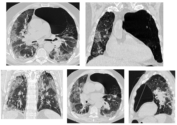
The naso-pharyngeal sampling was positive for SARS-CoV-2. So a chest CT (Picture 1 a-e) scan was urgently performed, in basal conditions, with a 64-slices multidetector scanner, and the images so obtained were analyzed with a slice-thickness of 1.2 mm and MPR reconstructions (axial, sagittal, and coronal).

Figure 1 (a-e): Chest X-ray showed the bilateral Covid-19 pneumonia signs, and the presence of a large, voluminous, bulla of pulmonary emphysema (15 cms) in the upper lobe of the left lung.
CT had documented in both lungs the presence of multiple areas with “ground glass” pattern, with interstitial thickenings, and other bilateral consolidations areas, as Covid-19 bilateral pneumonia. But CT also demonstrated the presence of bilateral emphysema, with the present of a large, voluminous, bulla (15 cms) of pulmonary emphysema occupying the upper lobe of the left lung. There were not pleural-pericardial effuses.
The patient was immediately hospitalized and treated. After two weeks the nasopharyngeal sampling had become negative for SARS-CoV-2, and so he continued therapy back at home, and periodical imaging follow-up had been programmed.
Discussion
Several studies in the literature had reported parenchymal findings of Covid-19 pneumonia, that are: bilateral, peripheral, and basal predominant GGO; crazy-paving pattern; consolidations; nodules; reticulations; interlobular septal thickenings; linear opacities; subpleural curvilinear lines; bronchial wall thickenings, often with an extensive geographical distribution. Unenhanced chest CT is useful in early diagnosis of Covid-19 infection, in monitoring disease progression, coinfection, or disease stability [4,5]. Chest CT can accurately evaluate the type and extent of lung lesions [6]. Chest High Resolution Computed Tomography can clearly show the radiological alterations resembling a pattern of combined pulmonary Covid-19 pneumonia and pulmonary emphysema
Conclusion
CT plays an important role in the diagnosis and severity evaluation of Covid-19 pneumonia, because it investigates very well the dynamic CT changes in different stages of the disease, and also in the follow-up of the patients. CT scan has been shown to be very important in detecting the radiological alterations resembling a pattern of combined pulmonary Covid-19 pneumonia and pulmonary emphysema. In general, CT played an important role in the diagnosis and evaluation of this global health emergency.
Conflicts of Interest: The authors certify that there is no conflict of interest with any financial organization regarding the material discussed in the manuscript.
Patient Consent Statement: The patient confirmed the condense for publication of our case report.
References
- Zu ZY, Jiang MD, Peng P, Chen W, Ni QQ, Lu GM. Coronavirus disease 2019 (Covid-19): a perspective from China. Radiology 2020; 296: E15–25.
- ACR recommendations for the use of chest radiography and computed tomography for suspected Covid-19 infection. https://www.acr.org/Advocacy-and-Economics/ACR-Position-Statements/Recommendations-for- Chest-Radiographyand-CT-for-Suspected-Covid-19-Infection.
- Jin Y-H, Cai L, Cheng Z-S, Cheng H, Deng T, Fan YP, Fang C, et al. A rapid advice guideline for the diagnosis and treatment of 2019 novel coronavirus (2019-nCoV) infected pneumonia (standard version). Mil Med Res. 2020; 7(1): 4.
- Kanne JP, Little BP, Chung JH, Elicker BM, Ketai LH. Essentials for radiologists on COVID- 19: an update. Radiology Scientific Expert Panel. Radiology 2020.
- Bernheim A, Mei X, Huang M, Yang Y, Fayad ZA, Zhang N, et al. Chest CT findings in coronavirus disease-19 (COVID-19): relationship to duration of infection. Radiology 2020.
- Nasir MU, Roberts J, Muller NL, Macri F, Mohammed MF et al. The role of emergency radiology in Covid-19: from preparedness to diagnosis. Can Assoc Radiol J 2020; 71(3): 293–300.

