Wilkie syndrome: Case Report
BEN ELHEND Salah*, DOULHOUSSNE Hassan, FASSI FIHRI Jawad, ROUKHSI redouane, ELFIKRI Abdelghani
Departement of Radiology, 5th Military Hospital, Guelmim, Morocco
Departement of Surgery, 5th Military Hospital, Guelmim, Morocco
Received Date: 15/04/2021; Published Date: 28/04/2021
*Corresponding author: BEN ELHEND Salah, Departement of Radiology, 5th Military Hospital, Guelmim, Morocco. Email: salahbenel4000@gmail.com, phone : +212 677 27 70 53
Abstract
Wilkie syndrome or superior mesenteric artery syndrome (SMAS) or cast syndrome is a rare condition caused by intermittent obstruction of the third portion of the duodenum compressed between the Superior Mesenteric Artery (SAM) anteriorly and the aorta and lumbar spine posteriorly.
It is a pathology that is often seen in children and young adults, and in cases of severe undernutrition. there are no specific clinical or biological criteria. CT scan, coupled with the ingestion of a radiopaque product or a gastroduodenal transit generally allows the diagnosis.
The management of this condition varies between observation, with correction of hydroelectrolytic disorders and the early initiation of nutrition, and surgery, depending on each particular case.
We aim to analyze the means of diagnosis and management of Wilkie syndrome in a pediatric population.
Keywords: Wilkie syndrome; Duodenum; Superior mesenteric artery; Computed Tomography
Introduction
Wilkie syndrome is an uncommon condition resulting from an extrinsic compression of the third portion of the duodenum compressed between the SAM anteriorly and the aorta and lumbar spine posteriorly. First described by von Rokitansky in 1842, SMAS was associated with orthopedic casting by Willet in 1878. Wilkie in 1927 formally characterized SMAS in a series of 75 patients [1,2].
SMAS usually presents more acutely than chronically with symptoms of small bowel obstruction and contrast-enhanced spiral computed tomography scan, coupled with the ingestion of a radiopaque product or a gastroduodenal transit generally allows the diagnosis. the evolution is fatal in absence of adequate management.
Case Report
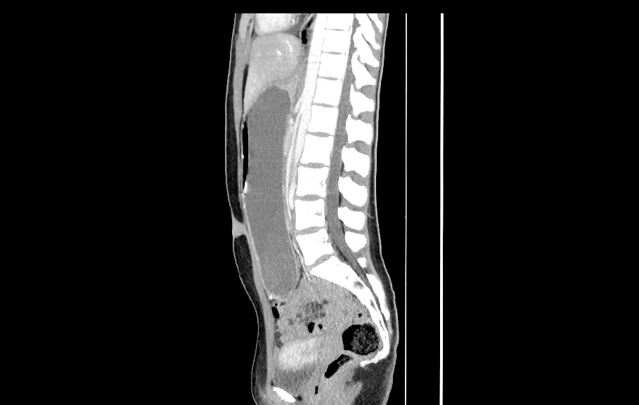
A 12-year-old girl, with history of chronic intermittent abdominal pain, vomiting and gastrointestinal problems apart from a weight-loss of approximately 6 kg over the last 4 months. It was only on the evening was admitted to emergency department for acute abdomen and vomiting. An abdominal CT scan showed a large gastric distension with the stenosis at the level of part III of the duodenum, and a reduced aorto mesenteric angle.
First, she was managed by using a nasogastric tube and with correction of hydroelectrolytic disorders, then a laparoscopic approach was performed to restore digestive continuity.


c
Figure: CT images in coronal (a) section showing a massive gastric distension, sagittal (b) and axial (c) sections, showing SMA (red arrow) exiting at an angle less than 13° from the aorta.
Discussion
SMAS is a rare form of upper gastrointestinal obstruction with a reported incidence of 0.013% to 0.3% [3].
Clinical findings include chronic intermittent abdominal pain, vomiting, nausea, early satiety and anorexia. The diagnosis of SMAS is commonly confirmed by upper gastrointestinal radiography but the diagnosis may be made by computerized tomography or laparotomy [4].
Endoscopic ultrasound at the site of duodenal compression has proven to be of substantial value, but angiography or angio-MRI are the best technique for determination of the aortomesenteric angle.
CT scan calculate the angle between the SMA and the aorta which is reduced from 7 ° to 22 °, whereas it is normally between 25 ° and 60 °. The aorto-mesenteric distance is also reduced and measures between 2 -8 mm, while the normal distance is 10 to 28mm [5]. In our patient the angle between the AMS and the aorta calculated on the CT images was 13°.
Surgical treatment is indicated in case of failure of medical treatment. It consists of a bypass by gastrojejunostomy or duodeno-jejunostomy, which can be performed laparoscopically [6] or modifying the anatomical conditions by mobilizing and uncrossing the duodeno-jejunal angl by positioning the jejunum to the right of the SMA after section of the Treitz muscle according to Strong's method [7].
Conclusion
Wilkie syndrome is an uncommon condition which should be suspected in case of small bowel obstruction symptoms and scan. Conditioning of the patient must begin before confirmation of the diagnosis by contrast-enhanced spiral computed tomography CT scan, angiography or angio-MRI.
Conflicts of Interest
The authors report no conflicts of interest.
References
- Wilkie DPD. Chronic duodenal ileus. Am J Med Sci 1927;173: 643Y9.
- Rokitansky, Innere-Hernien Handbuch der pathologischen Anatomie, Third volume, Wien: Braumüller & Seidel, 1842; pp. 215–219.
- Baltazar U, Dunn J, Floresguerra C, et al. Superior mesenteric artery syndrome: an uncommon cause of intestinal obstruction. South Med J 2000; 93: 606 Y 8.
- Biank V, Werlin S. Superior mesenteric artery syndrome in children: a 20-year experience. J Pediatr Gastroenterol Nutr. 2006; 42(5): 522-525.
- Welsch T, Büchler MW, Kienle P. Recalling Superior Mesenteric Artery Syndrome. Digestive Surgery. 2007; 24(3):149-156.
- Welsch T, Büchler MW, Kienle P. Recalling Superior Mesenteric Artery Syndrome. Digestive Surgery. 2007; 24(3):149-156.
- karren frederick merrill. Superior Mesenteric Artery Syndrome Treatment & Management.

