
Email id: info@ijclinmedcasereports.com
Contribution of High-Resolution Vessel Wall MRI (HR-VWI) to Understanding the Mechanism of Ischemic Stroke: Three Cases of Intracranial Atherosclerosis, Two Suggestive of Branch Atheromatous Disease
Khattab H¹, Mouddane M¹,*, Taoussi R2, Sikkal A1, Haddouali K1, Bellakhdar1, El Otmani H1, El Moutawakil B1 and Rafai MA¹
¹Department of Neurology, CHU Ibn Rochd, Casablanca, Morocco
²Department of Radiology, NAKHIL Radiology Center, Rabat, Morocco
Received Date: 23/10/2026; Published Date: 21/12/2026
*Corresponding author: Mouddane Meryem, Department of Neurology, CHU Ibn Rochd, Casablanca, Morocco
Abstract
Branch Atheromatous Disease (BAD) is an under-recognized cause of deep cerebral infarction, lying at the interface between cerebral small vessel disease and intracranial large-artery atherosclerosis. Its diagnosis is challenging: conventional MR angiography and CT angiography are frequently normal or inconclusive, as the culprit perforating arteries are too small to be visualized directly. We report three cases in which high-resolution vessel wall imaging (HR-VWI) helped clarify the mechanism of infarction. The first two patients presented with deep infarction and early neurological deterioration despite appropriately administered dual antiplatelet therapy, in the territory of the basilar artery perforators (Case 1) and in the lenticulostriate territory of the left middle cerebral artery (MCA) (Case 2). HR-VWI demonstrated an atheromatous plaque in the wall of the parent artery, with marked contrast enhancement in Case 2, confirming an atherothrombotic mechanism and leading to etiological reclassification (TOAST: large-artery atherosclerosis). The third patient presented with multiple infarcts in the vertebrobasilar territory; HR-VWI revealed multifocal stenoses of the posterior circulation (right vertebral artery V4 segment, left P2 segment). Intravenous tirofiban was used in the acute phase in all three patients. These cases illustrate the value of HR-VWI in elucidating the mechanism of infarcts with a normal or inconclusive conventional angiographic work-up, with implications for etiological classification, the intensity of antithrombotic and lipid-lowering therapy, and follow-up.
Keywords: Branch atheromatous disease; Plaque MRI; High-resolution vessel wall imaging (HR-VWI); Ischemic stroke; Basilar artery; Middle cerebral artery; Tirofiban
Introduction
Branch Atheromatous Disease (BAD) refers to deep infarction caused by an atheromatous microplaque at the origin of a perforating artery—the lenticulostriate arteries for the middle cerebral artery, the paramedian perforators for the basilar artery—as opposed to the distal lipohyalinosis typical of small vessel disease [1]. First described by Caplan in 1989 [1], BAD is clinically distinguished by a high risk of Early Neurological Deterioration (END) and progressive motor deficit, with END rates reaching up to 40% despite appropriately administered Dual Antiplatelet Therapy (DAPT), as the underlying pathophysiology—progressive microthrombosis within the perforating artery responds poorly to oral platelet inhibition alone.
The positive diagnosis of BAD remains difficult in routine practice: conventional MR angiography or CT angiography is most often normal, because the spatial resolution of these techniques does not allow direct visualization of a plaque located at the origin of a perforating artery less than one millimeter in diameter. Diagnosis therefore relies mainly on indirect evidence: infarct topography, clinical course (fluctuating or progressive), and exclusion of other causes. High-Resolution Vessel Wall Imaging (HR-VWI) may offer a decisive contribution in this setting: by directly imaging the wall of the parent artery (basilar artery, M1 segment), it can reveal a culprit mural plaque, with or without significant luminal stenosis, and characterize its degree of inflammatory activity through contrast enhancement.
We report three cases illustrating the contribution of this imaging technique to understanding the mechanism of ischemic stroke: two deep infarcts with an unfavorable course suggestive of BAD, one in the basilar artery territory and the other in the lenticulostriate territory of the middle cerebral artery, and one case of multiple vertebrobasilar infarcts revealing multifocal stenoses. We discuss the implications of these diagnoses for acute-phase management and secondary prevention.
Case 1: Basilar artery branch atheromatous disease
A 64-year-old man, whose only known vascular risk factor was hypertension, presented on March 27, 2026, with a left paramedian pontine ischemic stroke. Initial MRI showed a left paramedian pontine hyperintensity on Diffusion-Weighted Imaging (DWI) (Figure 1), with no significant stenosis on conventional MR angiography of the vertebrobasilar system. The patient was treated with intravenous thrombolysis followed by dual antiplatelet therapy; the initial LDL cholesterol level was 1.91 g/L (4.94 mmol/L). The initial course was favorable, with a modified Rankin Scale (mRS) score of 1 at discharge.
On April 14, 2026, 18 days later, the patient awoke with worsening dysarthria and new-onset swallowing difficulties, without motor deficit (last known well at 11 p.m.; presentation at 8 a.m.). Blood pressure was 150/90 mmHg, the NIHSS score was 1 (isolated dysarthria), and the 5-sip swallowing test was positive. Follow-up brain MRI confirmed extension of the left paramedian pontine ischemic lesion (concordant DWI and FLAIR findings), while MR angiography of the vertebrobasilar system again showed no identifiable significant stenosis.
Given this presentation suggestive of early neurological deterioration due to probable branch atheromatous disease—a clinically fluctuating, angiographically negative pattern not prevented by ongoing dual antiplatelet therapy—intravenous tirofiban was initiated in the acute phase. High-resolution vessel wall imaging of the basilar artery (dedicated protocol: high-resolution TOF, axial black-blood T2- and T1-weighted sequences before and after gadolinium injection, and TOF/T1 fusion), performed at a later stage on May 20, 2026, demonstrated a mural atheromatous plaque of the basilar artery abutting the origin of the paramedian perforators supplying the infarcted territory (Figure 2). This finding led to reclassification of the stroke etiology as large-artery atherosclerosis (TOAST), justifying intensification of lipid-lowering therapy, optimization of vascular risk factor control, and structured follow-up.
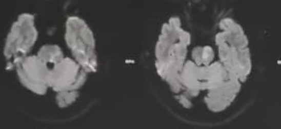
Figure 1: Initial diffusion-weighted MRI: left paramedian pontine DWI hyperintensities (axial slices).
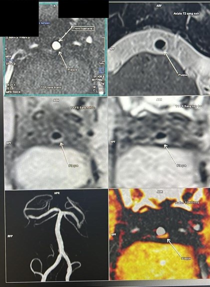
Figure 2: High-resolution vessel wall imaging (HR-VWI) of the basilar artery (May 20, 2026): mural plaque of the basilar artery abutting the origin of the paramedian perforators (arrows, “Plaque”), on high-resolution TOF, black-blood T2- and pre-/post-gadolinium T1-weighted sequences, and TOF/T1 fusion.
Case 2: Middle cerebral artery branch atheromatous disease
A 44-year-old overweight man presented on November 23, 2025, with transient sensory disturbance of the right hand associated with clumsiness. Initial diffusion-weighted MRI showed hyperintensities in the left Middle Cerebral Artery (MCA) territory (Figure 3A), and MR angiography suggested focal involvement of the left M1 segment (Figure 4).
The patient was hospitalized for 48 hours and treated with dual antiplatelet therapy (aspirin–ticagrelor) and high-intensity atorvastatin. The etiological work-up showed normal CT angiography of the supra-aortic arteries, an LDL cholesterol level of 0.4 g/L (1.03 mmol/L), and an HbA1c of 5.8%. Transcranial Doppler (TCD) showed a markedly elevated velocity in the left MCA (520 cm/s), suggestive of severe intracranial stenosis. Three weeks later, the patient’s condition worsened, and he also developed hematuria; follow-up diffusion-weighted MRI revealed new punctate ischemic lesions in the same left lenticulostriate territory (Figure 3B) despite ongoing dual antiplatelet therapy—a picture of early neurological deterioration due to probable branch atheromatous disease.
Intravenous tirofiban was administered for 48 hours, followed by resumption of dual antiplatelet therapy, intensified management of vascular risk factors, and close TCD monitoring. Antiplatelet therapy was adjusted (ticagrelor was replaced by clopidogrel because of hematuria). TCD monitoring showed fluctuating left MCA velocities during follow-up, with a downward trend at two months (approximately 350 cm/s). Intracranial angioplasty and stenting were considered in view of the persistent intracranial stenosis, but no final decision had been made at the time of writing.
High-resolution vessel wall imaging of the left M1 segment, performed on June 23, 2026 (high-resolution TOF protocol with black-blood T2- and T1-weighted sequences before and after gadolinium), confirmed a left M1 stenosis related to a mural plaque, with marked enhancement on post-gadolinium T1-weighted sequences (Figure 5)—a recognized marker of inflammatory activity and plaque vulnerability, and an independent risk factor for recurrence. This finding confirmed the atherothrombotic mechanism and led to reclassification of the etiology as large-artery atherosclerosis (TOAST).
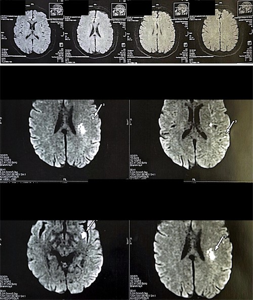
Figure 3: Initial diffusion-weighted MRI on November 24, 2025 (A, top) and follow-up at ~3 weeks (B, bottom): new punctate ischemic lesions in the left lenticulostriate territory (numbered arrows) despite ongoing dual antiplatelet therapy.
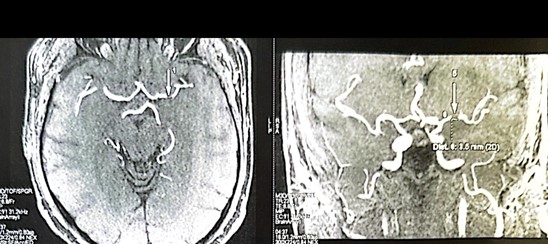
Figure 4: MR angiography (TOF): focal stenosis of the left M1 segment, with measurement of the residual lumen on the magnified reconstruction.
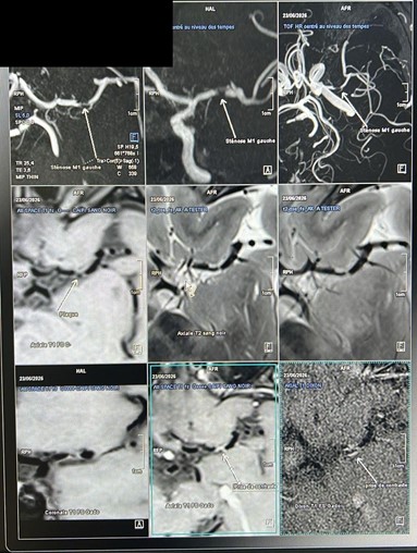
Figure 5: High-resolution vessel wall imaging (HR-VWI) of the left M1 segment (June 23, 2026): left M1 stenosis related to a mural plaque, visible on the three TOF projections (top row) and on black-blood T1- and T2-weighted sequences (middle row), with marked mural enhancement on post-gadolinium fat-suppressed T1 and Dixon sequences (bottom row)—a marker of plaque vulnerability.
Case 3: Multifocal intracranial atherosclerosis of the vertebrobasilar system
A 67-year-old man was admitted for sudden-onset imbalance associated with severe rotatory vertigo. Initial neurological examination revealed ataxia, with an Expanded NIHSS (e-NIHSS) score of 1.
Initial brain MRI showed multiple ischemic lesions in the vertebrobasilar territory, notably involving the territories of the Anterior Inferior Cerebellar Arteries (AICA) and posterior cerebral arteries (PCA) bilaterally (Figure 6).
Given the multifocal nature of the ischemic lesions and the risk of early recurrence, the patient was initially treated with intravenous tirofiban, followed by dual antiplatelet therapy combined with high-intensity statin therapy. Laboratory work-up showed an LDL cholesterol level of 0.76 g/L (1.97 mmol/L) and an HbA1c of 5.9%.
High-resolution vessel wall imaging of the posterior circulation (protocol combining high-resolution TOF and black-blood T1- and T2-weighted sequences before and after gadolinium injection) revealed multifocal stenoses, notably involving the V4 segment of the right vertebral artery and the P2 segment of the left posterior cerebral artery (Figure 7).
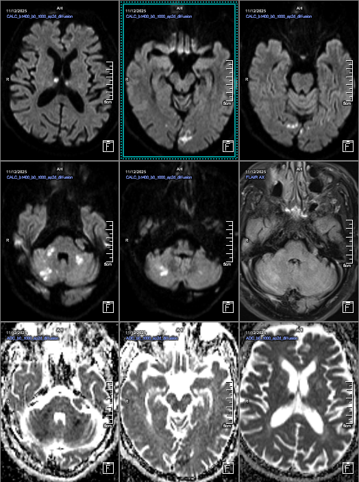
Figure 6: Axial brain MRI (diffusion-weighted imaging, ADC map, and T2-FLAIR): ischemic lesions in the bilateral PCA, AICA, and PICA territories.
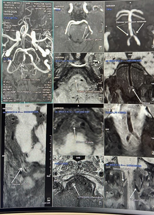
Figure 7: MR angiography and intracranial vessel wall imaging: multifocal stenoses of the posterior circulation, notably involving the V4 segment of the right vertebral artery and the P2 segment of the left posterior cerebral artery.
Discussion
BAD: a challenging diagnosis on conventional imaging
The concept of branch atheromatous disease, described by Caplan in 1989 [1], remains largely underdiagnosed in routine practice. The culprit plaque lies at the very origin of the perforating artery—too small to be resolved by conventional MR or CT angiography—so these examinations are most often reported as normal, wrongly suggesting simple small vessel lipohyalinosis. In our first case, initial MR angiography showed no significant stenosis, and it was the clinical course—early neurological deterioration despite appropriately administered dual antiplatelet therapy—that raised the hypothesis of BAD. In the second case, MR angiography and transcranial Doppler showed a focal M1 stenosis; HR-VWI nevertheless established its atheromatous nature, characterized its activity (contrast enhancement), and linked it to the infarcted lenticulostriate territory. It should be emphasized that, in this case, the presence of significant stenosis of the parent artery brings the presentation closer to a large-artery atherosclerotic mechanism with involvement of the perforator origin than to BAD in the strict sense, depending on the definitions used; the boundary between these entities remains debated.
Contribution of high-resolution vessel wall imaging (HR-VWI)
High-resolution vessel wall imaging overcomes this limitation by directly imaging the wall of the parent artery rather than its lumen alone. Khan et al. recently showed that HR-VWI could differentiate BAD from small vessel disease by demonstrating a mural plaque abutting the origin of the perforator, with contrast enhancement observed in approximately half of patients with confirmed BAD [2]. Such mural enhancement is considered a marker of inflammatory activity and plaque vulnerability: Zhao et al. showed that it was an independent predictor of ischemic recurrence in patients with intracranial large-artery atherosclerosis [3]. In our second case, the marked enhancement of the left M1 plaque is consistent with these data and, had this information been available earlier, might have strengthened the rationale for more intensive monitoring and treatment from the initial phase.
In Cases 1 and 2, HR-VWI confirmation was obtained only after the acute episode (approximately two and seven months after the index event, respectively) and therefore did not directly guide therapeutic decision-making in the hyperacute phase, which relied solely on the clinical evidence and conventional imaging. It did, however, retrospectively confirm the suspected atherothrombotic mechanism, allow reclassification of the etiology according to TOAST, and prompt corresponding adjustment of the intensity of secondary prevention. This argues for earlier integration of HR-VWI into the etiological work-up of deep infarcts with a clinical course suggestive of BAD, wherever the technique is accessible, rather than its use as a delayed second-line examination.
Our third case illustrates another contribution of HR-VWI: in the setting of multiple vertebrobasilar infarcts, the demonstration of multifocal stenoses (right V4, left P2) points toward diffuse intracranial atherosclerosis of the posterior circulation, which may influence the intensity of secondary prevention. This case, which does not represent BAD, highlights the value of the technique in distinguishing the mechanisms underlying multifocal infarcts.
Early neurological deterioration and the role of tirofiban
BAD is associated with a high risk of early neurological deterioration, estimated at up to 40% of cases, because oral dual antiplatelet therapy does not effectively block the final common pathway of platelet aggregation (fibrinogen binding to the glycoprotein IIb/IIIa receptor) that drives progressive microthrombosis within the perforating artery [4]. In a recent analysis combining propensity score matching and multivariable regression, Wen et al. showed that intravenous tirofiban significantly reduced the risk of END compared with oral antiplatelet therapy alone [4]. In addition, a post hoc analysis of the TREND trial suggested that the benefit of tirofiban was particularly marked in patients with severe intracranial stenosis [5], which matches the profile of the patient in Case 2 (severe M1 stenosis). The ongoing multicenter randomized TiMIS trial should help define the role of tirofiban followed by sequential dual antiplatelet therapy in mild-to-moderate ischemic stroke [6].
These data must nevertheless be weighed against the bleeding risk of tirofiban, which has been reported to be increased in some studies, although no significant increase in symptomatic intracranial hemorrhage was observed in the TREND trial. In our second case, the occurrence of hematuria on dual antiplatelet therapy (ticagrelor)—although not attributable to tirofiban itself—illustrates the need for close bleeding surveillance and individualized adjustment of long-term antithrombotic therapy.
Implications for management and limitations
Beyond the acute phase, identification of an atheromatous plaque in the parent artery modifies the secondary prevention strategy: TOAST reclassification as large-artery atherosclerosis, intensification of lipid-lowering therapy, optimization of vascular risk factor control, and, for severe symptomatic stenosis despite optimal medical therapy, discussion of intracranial angioplasty and stenting—an issue raised but not resolved in our second case.
This work has limitations inherent to the case report format: it describes three isolated cases interpreted descriptively, without histopathological confirmation of the plaque, and plaque imaging was performed only after the acute episode in Cases 1 and 2, which limits the conclusions that can be drawn about the contribution of HR-VWI to therapeutic decision-making in the hyperacute phase itself. Larger prospective studies incorporating HR-VWI into the initial etiological work-up are needed to establish its precise place in the diagnostic and therapeutic algorithm for deep infarcts suggestive of BAD.
Conclusion
These three cases illustrate the contribution of high-resolution vessel wall imaging to elucidating the mechanism of cerebral infarcts whose etiology remains uncertain after conventional work-up: two deep infarcts with an unfavorable course suggestive of branch atheromatous disease (basilar artery; M1 segment of the middle cerebral artery) and one case of multifocal infarcts revealing multiple stenoses of the posterior circulation. By directly visualizing the mural plaque of the parent artery and, where present, demonstrating its enhancement, HR-VWI allows etiological reclassification and targeted adjustment of secondary prevention. Earlier use of this technique, from the initial work-up of infarcts with an atypical clinical course, warrants prospective evaluation.
Conflicts of Interest: The authors declare no conflicts of interest related to this work.
References
- Caplan LR. Intracranial branch atheromatous disease: a neglected, understudied, and underused concept. Neurology, 1989; 39(9): 1246.
- Khan F, Goldstein E, Lewis S, Boxerman JL, Furie K, Yaghi S. High-Resolution Vessel Wall Imaging Can Differentiate Between Branch Atheromatous Disease from Small Vessel Ischemic Disease. J Am Heart Assoc, 2026; 15(4): e047631.
- Zhao Y, Song P, Feng P, Yuan S, Wu H, Cui J, et al. Plaque enhancement predicts recurrence in acute ischemic stroke patients with large artery intracranial atherosclerosis. J Stroke Cerebrovasc Dis, 2023; 32(12): 107406.
- Wen Q, Zhao Y, Deng Y, Guan S, Zhang S, Zhou J, et al. Tirofiban for Preventing Early Neurological Deterioration in Acute Ischemic Stroke Within 48 Hours of Onset: Evidence from a Dual-Method Analysis Using Propensity Score Matching and Multivariable Regression. CNS Neurosci Ther, 2025; 31(12): e70718.
- Wang J, Qiao Y, Li S, Li C, Wu C, Wang P, et al. Effects of tirofiban in preventing neurological deterioration in acute ischemic stroke with intracranial artery stenosis: a post hoc analysis of the TREND Trial. Eur Stroke J, 2025; 10(3): 919-928.
- Xu J, Peng H, Zhu Y, Xu F, Zhu L, Yang J, et al. Tirofiban with sequential dual antiplatelet therapy in mild acute ischemic stroke (TiMIS): protocol for a multicenter, randomized controlled trial. Ann Med, 2026; 58(1): 2644698.