
Email id: info@ijclinmedcasereports.com
The CASCADE Protocol: An Anatomy-Guided Sequential Methodology for Full-Face Rejuvenation
Natalia Sukmanskaya1, Alessio Redaelli2* and Robert Cimielewsky3
¹Laboratoires Revitacare, Saint-Ouen-l’Aumône, France
²Visconti di Modrone medical center Milan, Private Practice, Milan, Italy
3Prime clinic, private practice, Warsaw, Pologne
Received Date: 29/10/2026; Published Date: 16/12/2026
*Corresponding author: Alessio Redaelli, Visconti di Modrone medical center Milan, Private Practice, Milan, Italy
Abstract
Background: Periocular skin-quality changes and medial facial contour deficits may remain prominent when lateral facial volume and support are satisfactory. The CASCADE Protocol proposes targeted treatment of these concerns in a defined sequence.
Objectives: To describe the anatomical rationale, patient selection, and four-step CASCADE methodology, together with preliminary clinical observations.
Materials and Methods: This expert-derived protocol was applied in a descriptive series of 13 women (mean age, 44 years; range, 34–67) with moderate facial aging, mild skin laxity, and preserved lateral facial support. Each patient underwent one treatment session comprising four sequential steps: intradermal treatment of lateral periorbital skin quality and fine wrinkles; correction of the tear trough and lid–cheek transition; treatment of the nasolabial folds; and correction of the marionette region. Perioral fine wrinkles were additionally treated within Step 1 in five patients (38.5%). Non-cross-linked hyaluronic acid with a nutrient complex (Cytocare S line) was used for skin-quality treatment, and cross-linked hyaluronic (Revitafill) acid gels for contour correction. Placement and volumes were individualized. Assessment at 30–45 days included the treating injector’s clinical evaluation, patient-reported aesthetic outcomes on a descriptive, unvalidated five-category scale, and adverse-event recording.
Results: The treating injector observed improved periocular skin texture in all patients and improved perioral texture and fine wrinkles in those treated in that region. Overall aesthetic outcomes were rated as good by 10 patients (76.9%) and excellent by three (23.1%). No adverse events were reported or observed during treatment or follow-up.
Discussion: The CASCADE Protocol offers a structured, anatomy-guided sequence for full-face rejuvenation in selected patients. It complements existing approaches by addressing specific regional concerns through individualized treatment planning. Preliminary observations suggest its potential clinical utility; however, larger prospective studies with validated outcome measures and longer follow-up are needed to establish its efficacy, safety, and reproducibility.
Conclusion: CASCADE provides a structured treatment-prioritization framework for selected patients with periocular and medial facial aging concerns and relatively preserved lateral support. These preliminary observations do not establish comparative efficacy or safety. Larger prospective studies with validated outcomes, independent assessment, and longer follow-up are required to evaluate clinical utility, durability, and the added value of the treatment sequence.
Keywords: Hyaluronic acid; Dermal fillers; Facial ageing; Treatment methodology; Facial anatomy; Full-face rejuvenation; Tear trough; Nasolabial fold; Medical lifting
Introduction
Injectable facial rejuvenation has progressively evolved from the correction of individual wrinkles and folds toward comprehensive treatment planning based on facial anatomy, proportions, and the interaction between adjacent regions. This transition has encouraged clinicians to assess the face as a whole and to consider how regional changes contribute to its overall appearance and perceived emotional expression [1,2].
Several methodologies have helped formalize this approach. The MD Codes introduced a structured system of anatomical injection sites, incorporating recommendations on treatment depth, delivery technique, and product volume within a broader assessment of facial emotional attributes.2 However, corrective aesthetic medicine cannot be fully reduced to a standardized treatment template: individual differences in anatomy, aging patterns, tissue characteristics, previous treatments, and patient expectations require clinical judgment beyond predefined injection maps. Standardized frameworks should therefore support, rather than replace, individualized assessment and treatment planning. The AART-HIT methodology further emphasized systematic assessment, anatomical understanding, product selection, and individualized combinations of aesthetic injectables [3]. Although different in scope, these frameworks contributed to the development of more organized and comprehensive treatment strategies.
Within this evolution, the Domino Effect approach, described by Sukmanskaya and Redaelli, proposed a sequential treatment strategy based on facial biomechanics, in which correction of selected supporting regions was intended to produce benefits in adjacent areas.4 This concept highlighted the potential contribution of upper and lateral facial support to the appearance of the medial and lower face [4,5]. However, subsequent clinical experience highlighted limitations of relying on this approach alone and the need to consider the risks of temporal treatment. In the authors’ clinical experience, cases of alopecia were observed following temporal injections; this complication has also been described in the published literature.6 These observations underscore the need for careful consideration of the risks associated with treatment in this region. Furthermore, in the authors’ experience, centrofacial improvements frequently fell short of clinical expectations when the underlying local deficits were not adequately addressed. This limitation was particularly apparent in patients with already satisfactory lateral facial volume and support, in whom further lateral treatment offered limited benefit for persistent central facial concerns.
These observations prompted a reconsideration of the role of indirect correction in centrofacial rejuvenation. Tear-trough hollowing, an irregular lid–cheek transition, nasolabial folds, and marionette lines may remain prominent despite treatment of supporting regions. Moreover, changes in periocular skin texture constitute a recurrent concern that is not adequately addressed by structural support alone. In selected patients, similar changes in the perioral region, including fine wrinkles, further contribute to an aged centrofacial appearance. Lateral support alone may therefore be insufficient when the principal aesthetic concerns arise from localized anatomical deficits and changes in skin quality. Increasing treatment in supporting regions may fail to provide a proportionate improvement in the areas that originally motivated the patient to seek treatment, particularly when those supporting regions show little need for correction.
This clinical limitation provides the rationale for the CASCADE Protocol, an expert-derived, anatomy-guided methodology that combines whole-face assessment with targeted treatment of four regions in a defined sequence: the lateral periorbital region, the tear trough and lid–cheek transition, the nasolabial fold, and the marionette region. In selected patients, treatment of perioral wrinkles may be incorporated into the first step without modifying the four-step structure. This sequence establishes an order of clinical priorities, while indications, product selection, injection technique, and treatment volumes remain individualized. The guiding principle is to address the anatomical contributors to visible aging while limiting unnecessary product use and preserving individual facial characteristics.
The protocol was designed primarily for patients with early to moderate signs of facial aging, typically aged approximately 35–55 years, who seek a fresher, more rested appearance while preserving their individual facial identity. Patient selection is guided by clinical findings rather than chronological age alone. The principal concerns addressed are (i) deterioration in periocular skin quality, including uneven texture and fine wrinkles; (ii) a visible tear trough and irregular lid–cheek transition associated with volume loss or redistribution; (iii) deepening of the nasolabial folds; and (iv) accentuation of the marionette region, sometimes accompanied by a downturned appearance of the oral commissures. The corresponding treatment objectives are to improve periocular skin texture, soften the lid–cheek transition, reduce the prominence of nasolabial and marionette folds, and restore an overall rested appearance. In selected patients, perioral fine wrinkles and textural changes may also be addressed within the first step.
The objective of this article is to describe the rationale, treatment sequence, and technical principles of the CASCADE Protocol as a practical clinical prioritization framework, together with preliminary descriptive observations in 13 patients. The methodology aims to achieve appreciable correction with conservative product volumes, potentially reducing treatment costs while emphasizing anatomical safety. Whether this approach reduces product requirements and costs or improves safety compared with existing strategies remains to be established. A prospective study is planned to evaluate clinical outcomes, patient satisfaction, product consumption, and adverse events; the present article describes the methodology and its clinical rationale rather than demonstrating comparative superiority.
Applied Anatomical Rationale
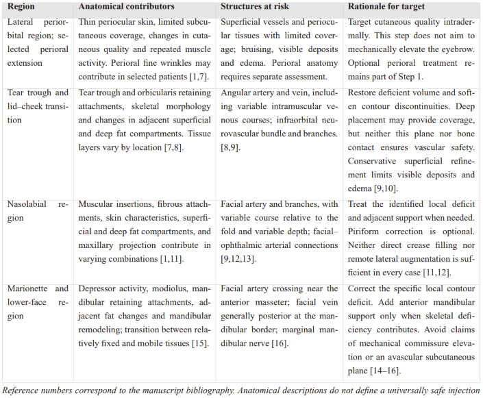
The overall anatomical basis for each step in the CASCADE Protocol is summarized in Table 1.
Table 1: Anatomical rationale and relevant structures by region.
Lateral periorbital region and tear trough
The lateral periorbital region and the tear trough represent distinct but complementary treatment targets. Thin periocular skin and limited subcutaneous tissue make fine wrinkles and textural irregularities particularly visible [1,7]. Accordingly, the first step targets skin quality rather than mechanical elevation or fixation of the lateral eyebrow. In selected patients, perioral fine wrinkles and textural changes may be addressed within the same step, with treatment adapted to the different tissue characteristics and muscular activity of that region.
The infraorbital region is characterized by thin skin, the orbicularis oculi muscle, and regionally variable superficial and deep soft-tissue compartments. It cannot be described uniformly as skin, muscle, and periosteum without intervening fat. Medially, the tear trough ligament anchors the tissues to the maxilla and continues laterally into the orbicularis retaining ligament. These attachments, together with skeletal morphology and changes in adjacent fat compartments, contribute to tear-trough hollowing and an irregular lid–cheek transition [7]. Correction therefore aims to restore deficient volume and soften contour discontinuities rather than release or reposition the retaining ligaments.
The angular vessels and infraorbital neurovascular bundle are important structures in treatment planning. Their position and depth vary, and the angular vein may follow an intramuscular course within the orbicularis oculi [8,9]. A deep-to-orbicularis approach may be appropriate for selected structural deficits, but neither this plane nor contact with bone guarantees avoidance of vascular injury. The infraorbital foramen and its emerging branches must be considered individually; a lateral entry point alone does not ensure that the neurovascular bundle will be avoided [9]. Conservative correction is particularly important because limited tissue coverage makes small contour irregularities, superficial deposits, and edema readily apparent [10].
Nasolabial region
The nasolabial fold reflects the interaction of muscular insertions, fibrous attachments, superficial and deep fat compartments, skin characteristics, and maxillary projection. Its prominence may therefore arise from different combinations of structural deficiency and tissue redistribution [1,11]. Treatment should address the anatomical contributors identified in each patient, which may include local volume deficits and deficient adjacent support, rather than assuming that either direct filling of the crease or remote lateral augmentation will be sufficient in every case.
The facial artery and its branches constitute a major safety consideration. Their relationship to the nasolabial fold is variable: the artery may pass medial or lateral to the fold and may change depth along its course [12]. Connections between the facial and ophthalmic arterial systems provide a potential route for filler-associated visual complications [9,13]. Consequently, no injection plane can be regarded as universally safe. Blunt cannula delivery, low injection pressure, and conservative volumes are precautions rather than guarantees against intravascular injection [9,14].
Marionette and lower-face region
Marionette lines reflect the combined effects of depressor muscle activity, the modiolus, mandibular retaining attachments, changes in adjacent fat compartments, and mandibular remodelling. The transition between relatively fixed tissues and more mobile tissues contributes to the visible fold and adjacent contour irregularities [15]. Correction should therefore be directed toward the specific local deficit rather than interpreted as mechanical elevation of the oral commissure.
The facial artery crosses the inferior mandibular border near the anterior edge of the masseter, with the facial vein generally posterior to it at this level. Their subsequent course and depth vary [16]. A superficial subcutaneous approach may be appropriate for selected marionette and prejowl deficits, but this plane is not avascular and does not eliminate the possibility of vascular injury [14,16]. These anatomical relationships support individualized assessment, controlled delivery, and conservative correction rather than reliance on a presumed safe plane.
Materials and Methods
The Four-Step CASCADE Treatment Methodology
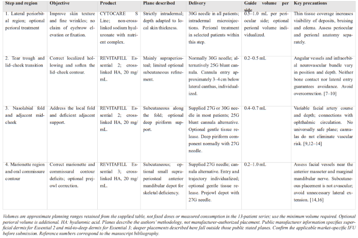
The four steps are performed in the sequence described below. Treatment volumes are individualized according to tissue characteristics, anatomical deficits, and the desired correction, using the minimum volume needed to achieve the intended endpoint throughout.
Step 1: Periocular skin quality and fine lines
The first step addresses skin texture and fine wrinkles in the lateral periorbital region. Its objective is to improve cutaneous quality rather than elevate or refixate the lateral eyebrow. Product is delivered intradermally, with placement depth adapted to local skin thickness, the selected formulation, and its intended use. In selected patients, treatment of perioral fine wrinkles and textural changes is incorporated into this step without altering the four-step sequence. A 30G needle was used in all patients.
Step 2: Tear trough and lid–cheek transition
The second step addresses infraorbital hollowing and aims to soften the lid–cheek transition. Any associated improvement in the upper nasolabial region is considered a possible secondary benefit rather than a predictable lifting effect.
Delivery is performed using a 30G needle or a 25G blunt cannula through a single entry point approximately three to four centimetres below the lateral canthus, adjusted to individual anatomy. The protocol uses a dual-plane approach when indicated. The supraperiosteal component is placed first and receives the larger proportion of the volume. A smaller subcutaneous component may then be delivered during withdrawal to refine residual contour irregularities. This superficial component is optional and requires particular restraint because thin tissue coverage increases the visibility of deposited product. Contact with bone does not, by itself, establish a safe injection plane [9,10]. A 30G needle is normally used; a 25G cannula may also be selected according to individual anatomy and operator experience.
Step 3: Nasolabial fold and adjacent mid-cheek
The third step addresses the nasolabial fold in relation to the adjacent mid-cheek and underlying support. In the majority of patients, treatment was performed using the supplied 27G needle or a 30G needle. A 25G blunt cannula may also be used, introduced through an entry point approximately one centimetre lateral to the oral commissure, with its position individualized to the planned trajectory.
Where indicated, gentle mechanical release of tethered tissues is performed, followed by conservative subcutaneous product placement along the fold. The deep piriform space is normally addressed with the supplied 27G needle. This component targets deficient deep support rather than the superficial crease alone and is not required in every patient.
Given the variable course and depth of the facial artery and its branches, both tissue release and product delivery require particular care [12]. Cannula use does not eliminate the risk of vascular injury or intravascular injection [14].
Step 4: Marionette region and oral commissure contour
The fourth step addresses marionette lines and contour deficiencies adjacent to the oral commissure. Product is delivered subcutaneously along the affected region through the supplied 27G needle. A cannula may alternatively be selected, with an entry point near the mandibular border and gentle mechanical release where indicated.
The entry point, trajectory, and extent of lateral treatment are adapted to individual anatomy, taking account of the facial vessels and the marginal mandibular branch of the facial nerve [16]. Correction is directed toward the identified contour deficit rather than mechanical repositioning of the oral commissure.
Where a prejowl deficiency includes a component of inadequate skeletal support, a small supraperiosteal depot may be placed on the anterior mandible, medial to the mandibular retaining attachment, before subcutaneous refinement. The supplied 27G needle is used for this indication. This deeper component is optional and depends on individual anatomy and expert clinical assessment.
Table 2: Summary of the four-step CASCADE methodology.
Product Selection and Rationale
Product selection follows the anatomy-guided principle underlying the CASCADE Protocol: each formulation is selected according to the local tissue characteristics and the specific treatment objective. The distinction is between improving skin quality and correcting structural volume deficits, with an emphasis on targeted treatment and conservative product use.
In the present description, three products from the Revitacare portfolio are used. CYTOCARE S Line, a non-cross-linked sodium hyaluronate solution containing a nutrient complex, is selected for the first step, which targets periocular skin texture and fine wrinkles. In selected patients, perioral textural changes and fine wrinkles may also be addressed within this step. Its intended role is to improve cutaneous quality rather than provide structural support or reposition the lateral eyebrow. This first step is performed strictly intradermally using a needle, whereas the subsequent steps involve needle and/or cannula delivery at different depths, according to the anatomical target and the structural deficit being addressed.
The subsequent steps use cross-linked hyaluronic acid gels to address localized volume and contour deficits. REVITAFILL Essential 2 is selected for the tear trough and lid–cheek transition (Step 2) and the nasolabial region (Step 3), whereas REVITAFILL Essential 3 is selected for the marionette region (Step 4). Both gels contain hyaluronic acid at 20 mg/mL, but their selection reflects differences in their intended placement and the requirements of the target tissues. Hyaluronic acid concentration alone does not establish equivalent mechanical behaviour or clinical suitability [17].
This selection is intended to support direct correction of the identified deficits while avoiding unnecessary augmentation of regions with already satisfactory volume and support. Product volumes remain individualized and are limited to those required to achieve the intended correction. The treatment sequence, products, delivery methods, approximate volume ranges, and key precautions are summarized in Table 2. The described injection planes must be distinguished from the manufacturer’s stated dermal indications; product-specific use should be checked against the applicable instructions for use. The principle of matching filler properties to tissue plane and mechanical demands is supported by the filler literature [17]. The products specified describe the formulations used in this protocol and do not establish superiority over alternative formulations.
Patient Population
The preliminary clinical series included 13 women with a mean age of 44 years (range, 34–67 years). All patients presented with moderate signs of facial aging and mild skin laxity, with clinical findings considered suitable for the targeted corrections described in the CASCADE Protocol. Their principal concerns involved age-related changes in the medial face, whereas lateral facial volume and support were considered clinically satisfactory and did not require additional treatment. Patient selection was based on clinical characteristics rather than chronological age alone.
Eligibility required no history of treatment with non-resorbable injectable materials and no aesthetic injectable treatment within the preceding six months. Additional exclusion criteria included active infection or inflammation in the intended treatment areas, known hypersensitivity to product components, and marked skin laxity considered unsuitable for correction with the proposed protocol.
Each patient underwent a single treatment session. In five of the 13 patients (38.5%), perioral fine wrinkles and textural changes also warranted treatment, which was incorporated into the first step without modifying the four-step sequence. Follow-up clinical assessment was performed 30–45 days after treatment.
Clinical outcomes during follow up period
The composite of TLF (target Lesion Failure) at 12± two months period was 3.8% with one cardiac death (0.95%),1 MI attributed to the target vessel (0.95%) and 2 TLRs solved by PCI. MACE rage during the follow up duration in brief were reported 7 (6.67%) patients consisting of: one cardiac death, one no cardiac death, one myocardial infarction attributed to the target vessel, one myocardial infarction non attributed to the target vessel (095%). TLR events were 2 patients (1.90%). All TLR events solved with PCI, with full recovery after treatment. TVR was observed in one patient (0.95%), solved by PCI. One incidence of ST was observed during the following period. No sub-acute ST was observed.no in hospital MACE and no late or very late thrombosis were observed.
Results
Preliminary outcomes were assessed between 30 and 45 days after a single treatment session in 13 women. Evaluation focused on patient-reported aesthetic outcomes, the treating injector’s clinical assessment, and treatment safety. These findings represent an initial descriptive assessment of the CASCADE Protocol.
The treating injector observed visible improvement in periocular skin texture in all 13 patients. Improvement in perioral skin texture and fine wrinkles was particularly apparent in the five patients (38.5%) who received additional treatment in this region during the first step.
Patient-reported overall aesthetic outcomes were classified into five descriptive ordinal categories: 1, worsened; 2, unchanged; 3, fair; 4, good; and 5, excellent. Outcomes were rated as good by 10 patients (76.9%) and excellent by three patients (23.1%). No patient reported a fair, unchanged, or worsened outcome. This classification was descriptive and did not constitute a validated patient-reported outcome measure.
No adverse events were reported or observed during treatment or throughout the 30–45-day follow-up period. These preliminary findings require evaluation in a larger prospective study with standardized outcome assessment and longer follow-up.
Discussion
Existing approaches to injectable facial rejuvenation differ in how they translate anatomical assessment into treatment priorities. The Domino concept emphasizes lateral structural support and its potential effects on adjacent facial regions; the MD Codes provide a detailed system of anatomical injection units and attribute-directed algorithms; and AART-HIT organizes assessment, anatomical reasoning, product selection, and individualized treatment planning [2-4]. CASCADE occupies a more focused position within this landscape: it proposes a defined regional sequence for selected patients with periocular and medial facial concerns, particularly when lateral support is relatively preserved. Its proposed contribution is therefore a practical treatment pathway for a specific presentation, rather than a comprehensive alternative to all existing approaches.
The Domino concept provides a biomechanical rationale for addressing lateral support before directly correcting medial signs of aging [4,5]. However, in the authors’ clinical experience, lateral correction alone may leave clinically relevant periocular fine lines, tear-trough hollowing, nasolabial folds, or marionette lines. Such residual concerns do not necessarily indicate inadequate lateral treatment; they may represent local changes requiring a different anatomical target or treatment modality. CASCADE addresses this clinical situation by organizing direct regional treatment when an indirect effect is insufficient or lateral augmentation is not the principal indication. The two approaches can therefore be complementary: lateral support should be addressed where deficient, while residual medial concerns require independent assessment rather than an assumption that further lateral volumization will resolve them.
The MD Codes offer a highly codified framework specifying injection sites, depths, delivery techniques, and suggested volumes. Their numerical structure provides explicit guidance, although the original publication emphasizes individualized code selection and adjustment of actual treatment volumes [2]. The relevant concern is consequently not codification itself, but an overly prescriptive application in which numerical targets take precedence over the clinical endpoint. CASCADE also standardizes aspects of treatment and is subject to the same risk. Its intended distinction is to organize decision-making around a limited sequence of regional objectives, with approximate volumes serving as guidance rather than mandatory doses. The appropriate endpoint remains the correction justified by the individual anatomy and treatment goal.
AART-HIT provides a broader framework, extending from whole-face assessment to product selection and an individualized treatment plan. Its priorities are derived from the patient’s presentation, whereas CASCADE fixes the order within a predefined clinical pathway.3 These frameworks operate at different levels: a comprehensive assessment may identify whether CASCADE is appropriate, while CASCADE specifies how a selected intervention is organized. Its narrower scope may facilitate teaching, documentation, and prospective evaluation, but also limits its applicability when the dominant deficit lies outside the four regions addressed. Standardization is useful only after appropriate patient selection; it cannot substitute for it.
Recent clinical evidence supports consideration of treatment sequence as a measurable variable. In a prospective split-face study of 15 patients, Hernandez and colleagues compared upper-face-first and lower-face-first injection sequences using the same amount and type of filler. Global Aesthetic Improvement Scale scores favored the upper-face-first sequence at the assessed time points, whereas differences in midfacial volume and jawline contour ratings did not reach statistical significance.18 These findings suggest that order may influence outcomes, but they do not validate the four-step CASCADE sequence or establish the appropriate timing of skin-quality treatment relative to volumetric correction. The specific sequence proposed here remains expert-derived and requires direct investigation.
A further consideration is the relationship between standardization and adaptability. Defined entry points, planes, product allocation, and approximate volumes make a methodology explicit and potentially easier to reproduce and audit. Nevertheless, these parameters require adjustment to individual anatomy, and landmark-based planning does not eliminate vascular risk [9,12-14,16]. The inclusion of both skin-quality treatment and structural correction also introduces multiple therapeutic components, whose individual contributions cannot be distinguished in the present report. Likewise, the specified products form part of the described intervention: equivalent outcomes with other formulations cannot be assumed, although this does not preclude future evaluation of the framework using alternative products [17].
The present article describes a methodology with preliminary observations in a small, uncontrolled series rather than establishing its comparative efficacy or safety. The sequence, injection planes, product allocation, and approximate volumes reflect clinical experience rather than formal consensus or controlled comparison. The 13-patient series, comprising women only, provides descriptive findings over 30–45 days, with an unvalidated patient rating and assessment by the treating injector. These features limit generalizability and introduce the possibility of selection and observer bias. The absence of observed adverse events in this sample cannot establish the frequency of uncommon complications. The findings do not establish superiority over Domino, MD Codes, AART-HIT, or individualized treatment without a fixed sequence. Moreover, the CASCADE sequence should be understood as a treatment-prioritization framework, not as a claim that facial aging follows the same chronological order. Prospective validation should include predefined eligibility criteria, a justified sample size, validated regional and global outcome measures, blinded photographic assessment, patient-reported outcomes, systematic adverse-event recording, and follow-up of six to twelve months. Comparative studies should distinguish the value of the treatment components from any additional benefit attributable to their sequence.
Conclusion
The CASCADE protocol proposes an anatomy-guided, four-step methodology for selected patients with periocular and medial facial aging concerns, particularly when lateral support is relatively preserved. Its intended contribution is a structured pathway integrating skin-quality treatment and regional correction while retaining individualized anatomical assessment and treatment endpoints. It may complement existing approaches when lateral support alone does not adequately address the patient’s concerns. The preliminary observations in 13 patients provide an initial clinical illustration; prospective comparative studies are required to determine its clinical utility, safety, durability, and the added value of the proposed sequence.
Funding: The source material on which this article is based was prepared for Revitacare for medical educational purposes.
Disclosure: The authors declare no conflict of interest.
Ethics and consent: This article describes a treatment methodology together with one patient treated in the course of routine clinical practice; formal institutional review board approval was therefore not sought. The patient provided written informed consent to treatment and to the publication of her photographs, and the work was conducted in accordance with the Declaration of Helsinki.
References
- Swift A, Liew S, Weinkle S, Garcia JK, Silberberg MB. The facial aging process from the “inside out.” Aesthet Surg J, 2021; 41(10): 1107–1119. doi:10.1093/asj/sjaa339.
- de Maio M. MD Codes™: A methodological approach to facial aesthetic treatment with injectable hyaluronic acid fillers. Aesthetic Plast Surg, 2021; 45(2): 690–709. doi:10.1007/s00266-020-01762-7.
- Nikolis A, Avelar LET, Haddad A, et al. Turn your AART into a HIT using a complete range of aesthetic injectables: Methodology for combining products to maximise patient outcomes. Clin Cosmet Investig Dermatol, 2024; 17: 2051–2069. doi:10.2147/CCID.S465155.
- Sukmanskaya N, Redaelli A. The Domino Effect: An innovative approach in aesthetic medicine leveraging the principles of facial biomechanics. J Biomed Res Environ Sci, 2024; 5(7): 868–871. doi:10.37871/jbres1965.
- Freytag L, Alfertshofer MG, Frank K, et al. Understanding facial aging through facial biomechanics: A clinically applicable guide for improved outcomes. Facial Plast Surg Clin North Am, 2022; 30(2): 125–133. doi:10.1016/j.fsc.2022.01.001.
- van den Elzen H, Schelke L, Velthuis PJ. Temporary hair loss after injection of hyaluronic acid filler. J Cosmet Dermatol, 2022; 21(2): 625–626. doi:10.1111/jocd.14453.
- Hong GW, Choi W, Yoon SE, Wan J, Yi KH. Anatomical-based filler injection diagnosis to treatment techniques: Infraorbital groove and hollowness. Life (Basel), 2025; 15(2): 237. doi:10.3390/life15020237.
- Calomeni M, Alfertshofer MG, Frank K, et al. Real-time ultrasound imaging of the tear trough: Lessons learned from functional anatomy. Aesthet Surg J, 2022; 42(5): 518–526. doi:10.1093/asj/sjab351.
- Vasconcelos-Berg R, Desyatnikova S, Bonavia P, Chammas MC, Navarini A, Sigrist R. Best practices for the use of high-frequency ultrasound to guide aesthetic filler injections—Part 2: Middle third of the face, nose, and tear troughs. Diagnostics (Basel), 2024; 14(22): 2544. doi:10.3390/diagnostics14222544.
- Trinh LN, Grond SE, Gupta A. Dermal fillers for tear trough rejuvenation: A systematic review. Facial Plast Surg, 2022; 38(3): 228–239. doi:10.1055/s-0041-1731348.
- Hong GW, Song S, Park SY, et al. Why do nasolabial folds appear? Exploring the anatomical perspectives and the role of thread-based interventions. Diagnostics (Basel), 2024; 14(7): 716. doi:10.3390/diagnostics14070716.
- Ten B, Kara T, Kaya Tİ, et al. Evaluation of facial artery course variations and depth by Doppler ultrasonography. J Cosmet Dermatol, 2021; 20(7): 2247–2258. doi:10.1111/jocd.13838.
- Doyon VC, Liu C, Fitzgerald R, et al. Update on blindness from filler: Review of prognostic factors, management approaches, and a century of published cases. Aesthet Surg J, 2024; 44(10): 1091–1104. doi:10.1093/asj/sjae091.
- Alam M, Kakar R, Dover JS, et al. Rates of vascular occlusion associated with using needles vs cannulas for filler injection. JAMA Dermatol, 2021; 157(2): 174–180. doi:10.1001/jamadermatol.2020.5102.
- Hong GW, Kim SB, Park SY, Wan J, Yi KH. Why do marionette lines appear? Exploring the anatomical perspectives and role of thread-based interventions. Skin Res Technol, 2024; 30(4): e13676. doi:10.1111/srt.13676.
- Vasconcelos-Berg R, Desyatnikova S, Bonavia P, Navarini A, Chammas MC, Sigrist R. Best practices for the use of high-frequency ultrasound to guide esthetic filler injections—Part 3: Lower third of the face. Diagnostics (Basel), 2025; 15(7): 921. doi:10.3390/diagnostics15070921.
- de la Guardia C, Virno A, Musumeci M, Bernardin A, Silberberg MB. Rheologic and physicochemical characteristics of hyaluronic acid fillers: Overview and relationship to product performance. Facial Plast Surg, 2022; 38(2): 116–123. doi:10.1055/s-0041-1741560.
- Hernandez CA, Uribe Zapata DE, Bermudez MP, et al. Clinical validation of the upper face first principle—A clinical, prospective, interventional split-face study. J Cosmet Dermatol, 2023; 22(2): 418–425. doi:10.1111/jocd.15563.