
Email id: info@ijclinmedcasereports.com
Synchronous Somatotroph Pituitary Neuroendocrine Tumor and BRAF V600E-Positive Mesial Temporal Ganglioglioma: Illustrative Case
Ramesh Chandra VV, Praneeth Prem kumar G*, Siva Prasad M, Prithvi Kanduri K, Rakesh Kumar K, Prasanna Kumar V, Venkata Krishna K and Bathuru Yashwanth
Department of Neurosurgery, SVIMS, Tirupati. Andhra Pradesh, India
Received Date: 18/07/2026; Published Date: 30/11/2026
*Corresponding author: Praneeth Prem kumar G, MS MCh, Assistant Professor, Department of Neurosurgery, SVIMS, Tirupati. Andhra Pradesh, India
Abstract
Background: Synchronous, spatially separate primary intracranial tumors of different histological lineages are uncommon. A functioning somatotroph pituitary neuroendocrine tumor (PitNET) occurring with a distant mesial temporal ganglioglioma is particularly unusual and creates diagnostic, pathological, and treatment-sequencing challenges.
Observations: A 43-year-old woman presented with a 1-year history of focal impaired-awareness seizures and a 6-month history of progressive acral and facial changes. Growth hormone was 34.4 ng/mL (laboratory reference range 0-3.6 ng/mL), and insulin-like growth factor 1 was 330 ng/mL; the age-adjusted reference interval was not available in the source record. MRI demonstrated a sellar macroadenoma with suprasellar extension and a separate left mesial temporal cystic-solid lesion. A staged strategy was used. Endoscopic endonasal transsphenoidal resection of the sellar tumor showed a PIT1- and growth hormone-positive, sparsely granulated somatotroph PitNET with a Ki-67 labeling index of 6%. Two months later, neuronavigation-guided minipterional resection of the temporal lesion showed a World Health Organization grade 1 ganglioglioma with BRAF V600E positivity and a Ki-67 index of 2%. At 3 months, growth hormone was 1.7 ng/mL and the patient was seizure-free (Engel class I) on antiseizure monotherapy.
Lessons: Atypical focal neurological symptoms in a patient with acromegaly should be investigated independently rather than attributed automatically to the sellar lesion. Staged resection can be a feasible individualized strategy when two symptomatic lesions require treatment. The coexistence of BRAF/MAPK activation in the ganglioglioma and systemic growth hormone/insulin-like growth factor 1 excess is hypothesis-generating, but a causal biological interaction cannot be inferred from a single case.
Keywords: Acromegaly; BRAF V600E; Case report; Ganglioglioma; Pituitary neuroendocrine tumor; Seizures
Abbreviations: GH = growth hormone; IGF-1 = insulin-like growth factor 1; IHC = immunohistochemistry; MRI = magnetic resonance imaging; PitNET = pituitary neuroendocrine tumor; WHO = World Health Organization
Background
Multiple primary intracranial tumors with different histological origins in the same patient are rare.1-3 This entity should be distinguished from a collision tumor, in which histologically different neoplasms are contiguous or intermixed at one anatomical site. Mixed sellar gangliocytoma-PitNET lesions have been reported,4 but they are biologically and anatomically different from a spatially separate temporal ganglioglioma coexisting with a functioning PitNET.
We describe a patient with acromegaly caused by a sparsely granulated somatotroph PitNET and drug-resistant focal seizures associated with a separate BRAF V600E-positive mesial temporal ganglioglioma. The report emphasizes diagnostic separation of the two syndromes, staged operative decision-making, and the limits of inferring molecular causality from a single observation.
Case
History and clinical evaluation:
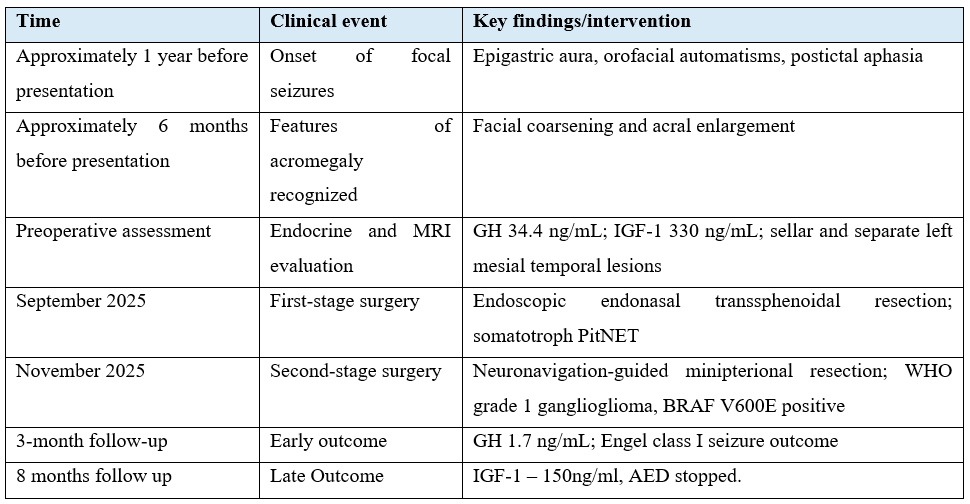
A 43-year-old woman presented with a 1-year history of recurrent focal impaired-awareness seizures. The episodes were described as an epigastric aura followed by orofacial automatisms and postictal aphasia, suggesting dominant mesial temporal onset. Seizure frequency increased despite antiseizure medication. Approximately 6 months after seizure onset, the patient and family noticed progressive coarsening of facial features and enlargement of the hands and feet.
The following clinically relevant information should be inserted from the source record, the patient is on Tab Levetiracetam 500 mg twice a day since the discharge after first surgery.
Endocrine and radiological assessment:
Basal GH was 34.4 ng/mL (laboratory reference range 0-3.6 ng/mL), and IGF-1 was 330 ng/mL. Because the age- and sex-adjusted IGF-1 reference interval or upper limit of normal was not provided, the degree of IGF-1 elevation cannot be standardized from the available documents. Pooled prolactin was 48.4 ng/mL; a 1:10 diluted sample measured 5.2 ng/mL. The dilution result argues against a high-dose hook effect, but it does not by itself establish stalk effect or exclude biological co-secretion. Cortisol was reported as 7.8 micrograms/dL. Visual field testing was normal.
MRI demonstrated two noncontiguous lesions: a sellar macroadenoma with suprasellar extension, described as Knosp grade 2, and a separate cystic-solid lesion in the left mesial temporal lobe that was T1 hypointense, T2 hyperintense, and heterogeneously enhancing.
Pre op MRI (25/7/2025) suggestive of an ill-defined, altered signal intensity lesion noted in the sella on the left side. Anterior pituitary gland on right side appears
homogeneously enhancing and seen displaced toward right side by the lesion. Superiorly, the lesion extends into the suprasellar cistern.
The infundibular stalk is seen displaced toward the right side. The optic chiasm is normal. Laterally on the left side, a lesion is seen extending into left cavernous sinus and encasing the cavernous segment of the left ICA (flow voids maintained). Posterolaterally, the lesion causes mass effect over left temporal lobe and effacement of the temporal horn of the left lateral ventricle; further the lesion extends into left ambient cistern and abuts the left cerebral peduncle.
The lesion is seen abutting /broad based towards the adjacent left tentorial dura. There is evidence of mild widening of the sella.
The lesion is heterogeneously isointense on T2 with few T2 hyperintense areas within,
isointense on T1 with few hypointense areas within. The lesion is showing heterogeneous enhancement after contrast administration.
The lesion is approximately 4 x 2.5 x 2.1 cm (AP x TR x CC).
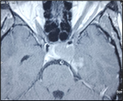
Figure 1: Axial Contrast image of the lesion.
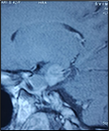
Figure 2: Sagittal contrast image.
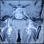
Figure 3: Coronal contrast image.
Post op MRI – 15/10/25 – Well-defined extra-axial altered signal intensity, which is isointense on T1, heterogeneously hyperintense on T2, not suppressed on FLAIR, showing focal area of blooming within, showing heterogeneous post-contrast enhancement measuring 1.9 x 1.5 x 1.2 cm noted in medial aspect of middle cranial fossa in parasellar location indenting medial temporal lobe, indenting left crus cerebri and effacement of temporal horn of left
lateral ventricle, left crural cistern.
Altered signal intensity noted in left half of sella and parasellar location which is hyperintense on T1 , hyperintense on T2 with few hypointense areas, T1 hyperintense areas are suppressed on FAT SAT FLAIR; showing blooming on SWI - could represent postprocedural changes.
Residual pituitary gland and stalk is displaced to the right side, with no obvious focal enhancing lesions appreciated within the pituitary gland – no residual tumor.
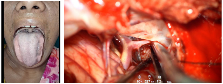
Image 1: Intra OP Images pterional craniotomy ( 2nd surgery).
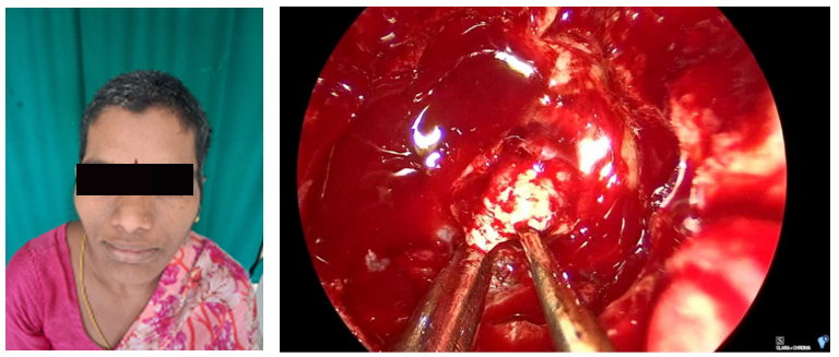
Image 2: Pre OP Images.
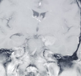
Figure 4: Coronal contrast image.
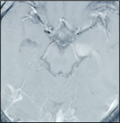
Figure 5: Axial contrast image.
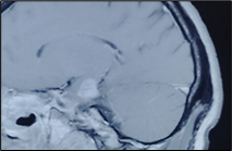
Figure 6: Sagittal contrast image.
Staged surgical management:
The multidisciplinary team selected staged treatment, addressing the functioning sellar tumor first and the epileptogenic temporal lesion second. This sequence was intended to obtain endocrine control and to separate the risks and goals of two different operations.
In September 2025, the patient underwent endoscopic endonasal transsphenoidal resection. The tumour was described intraoperatively as soft and vascular, and gross-total resection was achieved. Histology showed sheets of chromophobic cells. IHC was positive for PIT1 and GH. Pan-cytokeratin demonstrated fibrous bodies in fewer than 50% of cells, supporting a sparsely granulated somatotroph PitNET. The Ki-67 labelling index was 6%. Contemporary pituitary classification emphasises cell lineage, differentiation, invasion, and clinical behaviour rather than assigning aggressiveness from Ki-67 alone [5,5].
In November 2025, the patient underwent neuronavigation-guided left minipterional craniotomy and resection of the mesial temporal lesion. Histology showed neoplastic astrocytes and dysplastic neurons, including binucleated neurons. IHC was positive for GFAP, synaptophysin, and chromogranin. The lesion was diagnosed as a WHO grade 1 ganglioglioma.7 BRAF V600E positivity was reported, and the Ki-67 index was 2%.
NIMHANS Neuropathology – Lab Reference no - X – 5072/ 2025 & X – 4364 / 2025
Post Op CT suggest complete removal of the tumor in the temporal lobe.
HPE Images
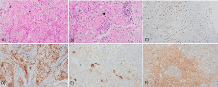
Figure 7: HPE images of Ganglioglioma.
A & B – 100 X & 200 X show dysplastic ganglionic cells (*) admixed with neoplastic Glial cells. Few eosinophilic granular bodies are seen (arrow)
C) OLIG – 2 IHC highlighting neoplastic cells.
D) Synaptophysin & E) Chromogranin labels dysplastic ganglionic cells.
F) BRAF p V 600E IHC is positive in tumor cells.
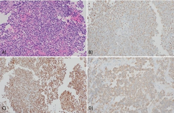
Figure 8: HPE image of Pituitary Neuroendocrine Tumour.
(A)HPE 200 X Neuroendocrine tumor with round monomorphic cells, stippled chromatin & amphophilic cytoplasm.
(B) Cytokeratin IHC showing perinuclear positivity
(C) PIT 1 transcription factor showing diffuse nuclear positivity
(D) GH IHC showing cytoplasmic positivity.
Follow-up and outcomes
Recovery after both operations was reported as uneventful. At 3-month follow-up, GH was 1.7 ng/mL(0.0 – 3.6) on 14/11/2025, and the patient remained seizure-free (Engel class I) on antiseizure monotherapy. Postoperative age-adjusted IGF-1: 150 ng/mL (50 – 300) on 23/1/2026
The last review of the patient is on 8/7/26, patient has no new symptoms and has stopped anti epileptics without seizure recurrence.
Timeline
Discussion
Observations
The principal observation is the synchronous presentation of two topographically separate and histologically distinct primary tumors: a functioning somatotroph PitNET and a mesial temporal ganglioglioma. Previous reports of multiple primary intracranial tumours include pituitary tumours coexisting with meningiomas, gliomas, and other neoplasms, but the overall phenomenon remains uncommon [1-3]. The present configuration is also distinct from a mixed sellar gangliocytoma-PitNET, in which neuronal and adenohypophyseal components occur together in the sellar lesion [4].
Gangliogliomas are low-grade glioneuronal tumors that commonly present with seizures. WHO classification recognises molecular alterations in the MAPK pathway, and BRAF V600E is found in a subset of gangliogliomas [7,9,10]. The temporal lesion therefore provides a plausible structural substrate for the patient's focal seizure semiology.
Biological hypothesis and its limits
GH and IGF-1 signalling can activate proliferative and survival pathways, including PI3K-AKT-mTOR, whereas BRAF V600E activates MAPK signalling [11,12]. Crosstalk between these pathways is biologically plausible. However, this case provides no serial growth curve, tissue-level receptor analysis, functional experiment, or evidence that the endocrine disorder preceded formation of the ganglioglioma. The coexistence should therefore be described as hypothesis-generating rather than as a demonstrated “double-hit” mechanism. Epidemiological observations of increased neoplasia in acromegaly do not establish the promotion of this specific tumor type [13].
Management considerations
Both lesions were symptomatic and surgically treatable. Treating the functioning PitNET first permitted early endocrine intervention and a separate assessment before epilepsy surgery. The alternative sequence, or a single-stage strategy, would depend on seizure urgency, mass effect, endocrine and anaesthetic risk, tumor anatomy, and institutional expertise. The favourable early outcome supports feasibility in this patient but does not establish superiority of the chosen sequence.
Limitations: This is a single case. There is no EEG data.
Lessons: Focal neurological symptoms in a patient with a pituitary disorder should be evaluated on their own merits, because a second pathology may coexist. Spatially separate tumors must not be mislabeled as a collision tumour. Staged surgical treatment can be effective when tailored to the dominant risks and goals of each lesion. Molecular pathway overlap is a useful research hypothesis, but causal language should be avoided without direct evidence.
Acknowledgments: This study is done with the help of Sri Balaji Arogya Vara Prasadini scheme.
Disclosures
Conflict of interest: None
Funding: Nil
Ethics: Applicable
Informed consent: Written informed consent for publication of the case and de-identified images was obtained from the patient.
References
- Furtado SV, Venkatesh PK, Ghosal N, Hegde AS. Coexisting intracranial tumors with pituitary adenomas: genetic association or coincidence? J Cancer Res Ther, 2010; 6(2): 221-223. doi:10.4103/0973-1482.65246.
- Tunthanathip T, Kanjanapradit K, Ratanalert S, Phuenpathom N, Oearsakul T. Multiple, primary brain tumors with diverse origins and different localizations: case series and review of the literature. J Neurosci Rural Pract, 2018; 9(4): 593-607. doi:10.4103/JNRP.JNRP_82_18.
- Amirjamshidi A, Mortazavi SA, Shirani M, Saeedinia S, Hanif H. Coexisting pituitary adenoma and suprasellar meningioma: a coincidence or causation effect? Iran J Neurosurg, 2017; 3(2): 64-69.
- Chen D, Xu J, Zhong P, Huang X, Xu M. Pituitary adenoma with gangliocytoma: report of two cases. Oncol Lett, 2014; 8(2): 781-784. doi:10.3892/ol.2014.2183.
- Asa SL, Mete O, Perry A, Osamura RY. Overview of the 2022 WHO classification of pituitary tumors. Endocr Pathol, 2022; 33(1): 6-26. doi:10.1007/s12022-022-09703-7.
- Trouillas J, Roy P, Sturm N, et al. A new prognostic clinicopathological classification of pituitary adenomas: a multicentric case-control study of 410 patients with 8 years post-operative follow-up. Acta Neuropathol, 2013; 126(1): 123-135. doi:10.1007/s00401-013-1084-y.
- Louis DN, Perry A, Wesseling P, et al. The 2021 WHO classification of tumors of the central nervous system: a summary. Neuro Oncol, 2021; 23(8): 1231-1251. doi:10.1093/neuonc/noab106.
- Giustina A, Barkan A, Beckers A, et al. Consensus on criteria for acromegaly diagnosis and remission. Pituitary, 2024; 27(1): 7-22. doi:10.1007/s11102-023-01360-1.
- Schindler G, Capper D, Meyer J, et al. Analysis of BRAF V600E mutation in 1,320 nervous system tumors reveals high mutation frequencies in pleomorphic xanthoastrocytoma, ganglioglioma and extra-cerebellar pilocytic astrocytoma. Acta Neuropathol, 2011; 121(3): 397-405. doi:10.1007/s00401-011-0802-6.
- Koelsche C, Wöhrer A, Jeibmann A, et al. Mutant BRAF V600E protein in ganglioglioma is predominantly expressed by neuronal tumor cells. Acta Neuropathol, 2013; 125(6): 891-900. doi:10.1007/s00401-013-1100-2.
- Pollak M. Insulin and insulin-like growth factor signalling in neoplasia. Nat Rev Cancer, 2008; 8(12): 915-928. doi:10.1038/nrc2536.
- Mendoza MC, Er EE, Blenis J. The Ras-ERK and PI3K-mTOR pathways: cross-talk and compensation. Trends Biochem Sci, 2011; 36(6): 320-328. doi:10.1016/j.tibs.2011.03.006.
- Dal J, Leisner MZ, Hermansen K, et al. Cancer incidence in patients with acromegaly: a cohort study and meta-analysis of the literature. J Clin Endocrinol Metab, 2018; 103(6): 2182-2188. doi:10.1210/jc.2017-02457.