
Email id: info@ijclinmedcasereports.com
Incidental Finding of Occult Metastatic Papillary Thyroid Carcinoma Within a Tuberculous Cervical Lymph Node and A Tumour-Free Thyroid: Diagnostic Dilemma of a Rare Association
Hajar El Agouri1,*, Mohamed Tbouda1, Achref Miry2, Marouane Balouki3 and Sanaa Abbaoui2
1Department of pathology Military Hospital Oued Eddahab, Agadir Morocco, Faculty of Medicine and Pharmacy, University Ibn Zohr, Agadir
2Department of pathology and laboratory medicine, Mohammed VI University Hospital, Faculty of Medicine and Pharmacy, University Ibn Zohr, Agadir
3Department of Otorhinolaryngology, Military Hospital Oued Eddahab, Agadir Morocco, Faculty of Medicine and Pharmacy, University Ibn Zohr, Agadir
Received Date: 03/10/2026; Published Date: 25/11/2026
*Corresponding author: Hajar El Agouri, Department of pathology Military Hospital Oued Eddahab, Agadir Morocco, Faculty of Medicine and Pharmacy, University Ibn Zohr, Agadir
Abstract
Background: Metastatic Papillary Thyroid Carcinoma (PTC) confined to cervical lymph nodes, with no identifiable primary tumour in a completely examined thyroidectomy specimen, is the rarest presentation of occult thyroid carcinoma, with fewer than 30 cases reported to date. Its coexistence with tuberculous lymphadenitis within the same lymph node has only rarely been observed.
Case presentation: A 45-year-old woman, with no relevant medical history, presented with a slowly enlarging right level II cervical mass of one year's duration, associated with a 4-kg weight loss over six months. Ultrasonography showed a heterogeneous, necrotic nodal mass suggestive of tuberculous lymphadenitis, with a normal-appearing thyroid gland. The nodal conglomerate was excised and entirely submitted for histological examination, which revealed caseating granulomatous lymphadenitis; Mycobacterium tuberculosis was detected by the GeneXpert MTB/RIF assay, without rifampicin resistance. Within the same lymph node, an 8-mm focus of metastatic PTC was identified, composed of infiltrative follicles set in a desmoplastic stroma and displaying the characteristic nuclear features; the tumour cells were positive for CK7, thyroglobulin and TTF-1 antibodies. A subsequent total thyroidectomy with bilateral neck dissection showed no carcinoma and no fibrous scar in the entirely embedded thyroid gland, and all 15 dissected lymph nodes were free of metastasis (pT0 N1b M0). The patient received nine months of antituberculous therapy without radioactive iodine and remained free of disease at one-year follow-up.
Conclusions: The absence of an intrathyroidal primary raises three hypotheses: an undetected microcarcinoma, a regressed primary, or malignant transformation of intranodal ectopic thyroid tissue. In tuberculosis-endemic regions, a diagnosis of caseating granulomatous lymphadenitis should not preclude complete histological examination of the lymph node, as both conditions share clinical and radiological features and may coexist within the same node.
Keywords: Papillary thyroid carcinoma; Occult papillary thyroid carcinoma; Lymph node metastasis; Tuberculosis; Lymphadenitis; Case report
Background
Papillary Thyroid Carcinoma (PTC) is the most frequent thyroid malignancy and spreads early to regional lymph nodes [1,2]. A cervical nodal metastasis is sometimes its first sign, and a small primary is then usually found in the thyroidectomy specimen. Much rarer is the situation in which no primary tumour is found despite complete histological study of the gland. Boucek et al. classified occult thyroid carcinoma into four groups [3], and Liu et al. later proposed a fifth group, in which metastases occur while the thyroid is benign on both imaging and pathology [4]. Xu et al. retrieved only seven such cases from a large institutional archive [5], and a pooled analysis published in 2026 counted 23 cases in total [6].
Tuberculous lymphadenitis is the most frequent form of extrapulmonary tuberculosis (TB) in Morocco, where it accounted for 41% of notified extrapulmonary cases in 2021 [7,8]. It mimics nodal metastasis of PTC clinically and radiologically [9,10]; the two diseases have been reported together in the same patient mainly as isolated cases [11–13], and within a single lymph node only exceptionally [11,14]. We report a patient in whom both conditions were found in a single cervical node while the thyroid gland was free of tumour, and we review both bodies of literature.
Case Presentation
History and physical examination:
A 45-year-old woman presented with a right cervical swelling in the upper jugular region, which had been slowly enlarging for one year. She had never received neck irradiation, had no family history of thyroid disease and reported no contact with tuberculosis. She had no fever but reported progressive weight loss of 4 kg over the previous six months. Physical examination revealed an upper jugular (level II) mass, which was mobile, firm, and painless, measuring approximately 6 cm on palpation. Thyroid gland palpation was unremarkable.
Investigations and initial surgery:
The chest radiograph was normal. Cervical ultrasound revealed a heterogeneous, necrotic, mobile mass, located in the upper jugular region (level II), primarily suggestive of tuberculous lymphadenopathy. The thyroid gland was normal, and the thyroid-stimulating hormone (TSH) level was also within the normal range. The nodal conglomerate was excised through a limited cervical incision in the otorhinolaryngology department and sent for histopathological examination.
Pathological findings:
The specimen consisted of a matted conglomerate of right upper jugular lymph nodes (level II). Its cut surface showed friable necrotic foci. The entire specimen was submitted for histological examination. Microscopically, the nodal architecture was largely effaced by geographic areas of eosinophilic, finely granular, acellular necrosis of caseous type, bordered by a rim of epithelioid histiocytes, Langhans-type giant cells, lymphocytes and fibroblasts; residual lymphoid parenchyma persisted at the periphery (Figure 1).
The GeneXpert MTB/RIF assay performed on the specimen detected Mycobacterium tuberculosis, with no evidence of rifampicin resistance.
Within the same lymph node as the granulomatous lesions, a focus of carcinoma measuring 8 mm in greatest dimension was present. It consisted of angulated, infiltrative follicles and tubules, with some papillary structures, containing dense colloid, set in a desmoplastic, inflamed stroma and in direct contact with residual lymphoid tissue (Figure 2). The tumour cells showed enlarged, oval, overlapping nuclei with cleared ("ground-glass") chromatin, irregular nuclear contours and nuclear grooves (Figure 3). There were no psammoma bodies, capsular breach or extranodal extension. On immunohistochemistry, the tumour cells were positive for cytokeratin 7 (CK7), thyroglobulin and thyroid transcription factor-1 (TTF-1), and negative for cytokeratin 20 (CK20) (Figure 4). The final diagnosis was metastatic papillary thyroid carcinoma associated with tuberculous lymphadenitis.
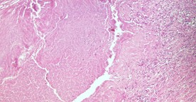
Figure 1: Tuberculous lymphadenitis: Interface between the granular eosinophilic caseous-type necrosis and a band of epithelioid histiocytes, Langhans-type giant cells, lymphocytes and fibroblasts (Hematoxylin-eosin stain, original magnification ×200).
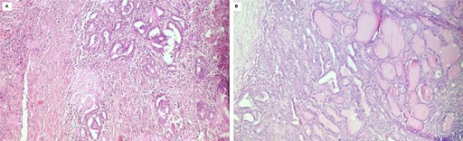
Figure 2: Incidental finding of occult metastatic papillary thyroid carcinoma in the same lymph node: (A) Neoplastic follicles and tubules infiltrating the nodal lymphoid tissue (Hematoxylin-eosin stain, original magnification ×200). (B) Follicular-patterned area with angulated infiltrative follicles, containing dense colloid, set in a desmoplastic stroma and in direct contact with residual lymphoid tissue (Hematoxylin-eosin stain, original magnification ×200).
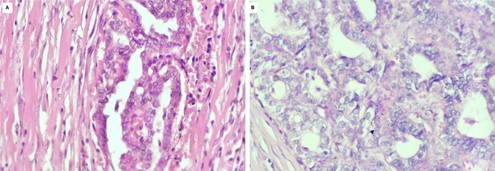
Figure 3: (A) and (B): Nuclear features of PTC at high power: Enlarged, crowded and overlapping nuclei with cleared chromatin, irregular contours and grooves (Hematoxylin-eosin stain, original magnification ×400).
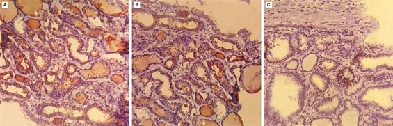
Figure 4: (A), (B) and (C): Immunohistochemistry of the nodal carcinoma: the tumour cells are positive for (A) CK7, (B) thyroglobulin and (C) TTF-1 (original magnification ×200).
Staging work-up and definitive surgery:
Cervical ultrasound revealed a thyroid gland of normal size and echostructure, with no nodules and no cervical lymphadenopathy. Thyroid function tests were within normal limits, including a normal TSH level and normal thyroglobulin levels. Following multidisciplinary team discussion, a total thyroidectomy with bilateral cervical lymph node dissection was subsequently performed as a second-stage procedure. The thyroid was fully embedded in 15 blocks. It showed normal parenchyma without carcinoma or microcarcinoma, and no fibrous scar that might mark a regressed tumour. All 15 lymph nodes from the bilateral dissection were free of metastasis. The disease was staged pT0 N1b M0 (AJCC 8th edition; stage I, age under 55 years).
Treatment and follow-up:
Anti-tuberculous treatment was started three months after the positive GeneXpert result. It was based on the standard Moroccan national regimen for new cases of lymph node tuberculosis: two months of rifampicin, isoniazid, pyrazinamide and ethambutol, then four months of rifampicin and isoniazid (2RHZE/4RH), with the continuation phase extended to give a total duration of nine months (2RHZE/7RH). After thyroidectomy, levothyroxine replacement was started at 1.5 µg/kg/day. Radioiodine was not administered, and postoperative thyroglobulin was not measured. One year after diagnosis, at the end of treatment, clinical examination and cervical ultrasound showed no evidence of recurrent carcinoma or tuberculosis.
Discussion
A rare association entity:
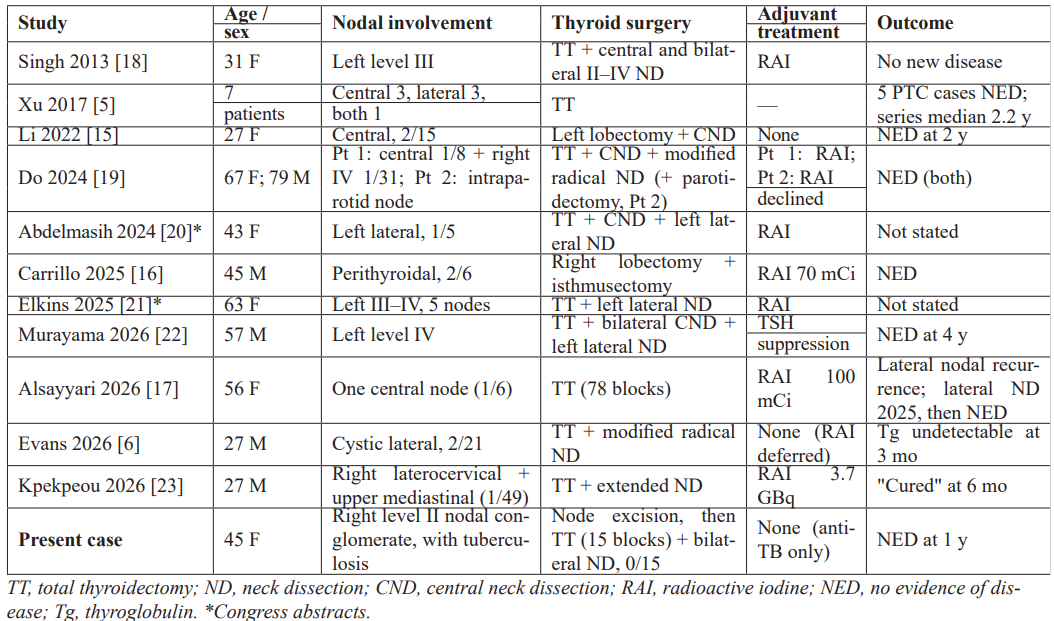
Nodal PTC with a tumour-free thyroid remains rare; Table 1 summarises the published cases in which thyroid surgery was performed. In the pooled analysis of 23 cases by Evans et al., the sex ratio was nearly equal, 65% of patients presented with a lateral neck mass, and 87% had no evidence of disease at a median follow-up of 24 months [6]; in the series of Xu et al., all five patients with PTC were alive without disease (median follow-up of the series, 2.2 years) [5]. The frequency of this entity is probably underestimated, as it depends on how completely the thyroid is examined: one published case had only a lobectomy [15], serial sectioning of the whole gland has been recommended [16], and nodal recurrence may appear years later [17]. Our patient fits this profile, with a lateral nodal deposit, a normal-appearing gland and a favourable early course.
Table 1: Published cases of nodal papillary thyroid carcinoma without an identifiable primary after thyroid surgery.
Where does the carcinoma come from?
Three mechanisms have been proposed. The first is an undetected intrathyroidal microcarcinoma: foci under 3 mm can escape routine histological sampling [15], and occult microcarcinomas are a recognised source of nodal metastasis [24]; this explanation is weakened, although not excluded, when the whole gland has been embedded, as in our patient. The second is a regressed primary: spontaneous regression has been documented in PTC, including in a nodal metastasis [25], but its histological footprint, a stellate fibrous scar, was absent from our patient's fully embedded gland, and was seen in only one of seven glands in the series of Xu et al. [5]. The third is carcinoma arising in intranodal ectopic thyroid tissue. Benign thyroid inclusions in cervical nodes have long been recognised [26,27], and PTC arising in intranodal or ectopic thyroid tissue with a normal gland has been reported [23,28,29]. Although inclusions are classically small, capsular and bland, they may show nuclear changes, whereas nuclear pseudoinclusions, desmoplasia and effacement of the nodal architecture favour metastasis [30]. The infiltrative, desmoplastic tumour in our case is therefore carcinoma, and a tumour-free thyroid with 15 negative nodes makes an intranodal origin plausible; however, our material does not show a transition from benign ectopic follicles, so this origin remains unproven. BRAF V600E immunohistochemistry or molecular testing could at least document a PTC-type driver [5,30].
Tuberculosis and papillary carcinoma in the same lymph node:
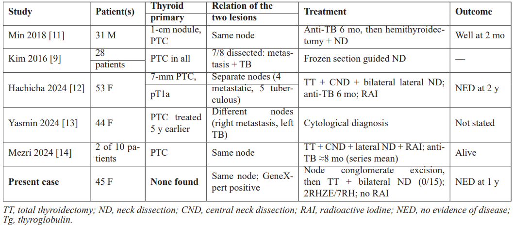
Tuberculous lymphadenitis and nodal metastasis of PTC share their preferred location in the lateral neck and several imaging features, including hypoechoic, necrotic or cystic nodes and calcifications [10,13]; in a Korean series of 9,098 operated patients, 28 had tuberculous lateral nodes initially read as metastases [9]. Coexistence within a single node has been documented only rarely, always with a known or synchronous thyroid primary [11,14]; more often, the two lesions involve separate nodes of the same neck [12,13,31–33]. Representative reports are summarised in Table 2. In our patient, by contrast, no thyroid primary was found. Whether the association is causal is unknown: tuberculosis is associated with an increased overall risk of cancer [34], and chronic mycobacterial inflammation has been proposed as a promoter of carcinogenesis [12,14], but in an endemic area simple coincidence is at least as likely.
Table 2: Reported coexistence of tuberculous lymphadenitis and metastatic papillary thyroid carcinoma.
Practical implications:
For the pathologist, a diagnosis of caseating lymphadenitis should not end the examination of a cervical node: the whole node should be embedded, since a metastatic deposit may occupy only a few millimetres of it; the tuberculous nature of the necrosis should be confirmed by Ziehl–Neelsen staining, molecular testing or culture [11,14], as GeneXpert did here; and immunohistochemistry (TTF-1, PAX8, thyroglobulin and BRAF V600E) should complement, not replace, morphology [30]. For management, total thyroidectomy is the accepted next step even when the thyroid appears normal, as it removes a possible ultramicroscopic primary and allows thyroglobulin-based surveillance [2,6,18], and lateral disease requires therapeutic neck dissection [2]. Radioiodine has been used in most reports [16–21,23,29], but it may be deferred in selected patients [6], and the 2025 American Thyroid Association guidelines base this decision on a four-tier risk of recurrence [2]; it was withheld in our patient, given a single 8-mm deposit without extranodal extension, a tumour-free thyroid and 15 negative lymph nodes. When tuberculosis coexists, anti-tuberculous treatment has usually been given around the time of surgery [12,14,32], and lymphadenopathy that persists under treatment should prompt a search for a coexisting malignancy [11,14].
Conclusion
This case adds to the small group of nodal papillary thyroid carcinomas with a tumour-free thyroid, and illustrates the rarely observed situation in which the metastatic deposit lies within a tuberculous lymph node. In tuberculosis-endemic regions, finding granulomas in a cervical node should not end the search for malignancy, and a normal thyroid on ultrasound does not exclude a thyroid origin. Total thyroidectomy remains the basis of management; the need for radioiodine should be weighed case by case, and long-term surveillance is required.
Declarations
Ethics approval: The manuscript complies with ethical publication standards and contains no identifying patient information.
Consent for publication: Written informed consent was obtained from the patient for the publication of this case report and the accompanying images.
Availability of data and materials: Not applicable.
Competing interests: The authors declare no competing interests.
Funding: This research received no specific grant from any funding agency in the public, commercial or not-for-profit sectors.
Authors' contributions and acknowledgements: All authors have participated in the preparation of the draft, have read and agreed to the published version of the manuscript.
References
- Baloch ZW, Asa SL, Barletta JA, et al. Overview of the 2022 WHO classification of thyroid Endocr Pathol, 2022; 33(1): 27–63. doi:10.1007/s12022-022-09707-3.
- Ringel MD, Sosa JA, Baloch Z, et al. 2025 American Thyroid Association management guidelines for adult patients with differentiated thyroid cancer. Thyroid, 2025; 35(8): 841–985. doi:10.1177/10507256251363120.
- Boucek J, Kastner J, Skrivan J, et al. Occult thyroid carcinoma. Acta Otorhinolaryngol Ital, 2009; 29(6): 296–304.
- Liu H, Lv L, Yang Occult thyroid carcinoma: a rare case report and review of literature. Int J Clin Exp Pathol, 2014; 7(8): 5210–5214.
- Xu B, Scognamiglio T, Cohen PR, et al. Metastatic thyroid carcinoma without identifiable primary tumor within the thyroid gland: a retrospective study of a rare phenomenon. Hum Pathol, 2017; 65: 133–139. doi: 10.1016/j.humpath.2017.05.013.
- Evans MJ, Sonnenburg AM, Elliott E, Otto AL, McIntire K. Occult papillary thyroid carcinoma with no identifiable intrathyroidal primary: a case report and pooled analysis of 23 published cases. Cureus, 2026; 18(7): e112518. doi:10.7759/cureus.112518.
- Ministère de la Santé et de la Protection Sociale, Programme National de Lutte Antituberculeuse. Lignes directrices nationales pour le diagnostic et la prise en charge de la tuberculose extrapulmonaire. Rabat: Ministère de la Santé et de la Protection Sociale, 2023.
- Hamzaoui G, Amro L, Sajiai H, et al. Lymph node tuberculosis: epidemiological, diagnostic and therapeutic aspects, about 357 cases. Pan Afr Med J, 2014; 19: 157. doi:10.11604/pamj.2014.19.157.4916.
- Kim SM, Jun HH, Chang HJ, et Tuberculosis cervical lymphadenopathy mimics lateral neck metastasis from papillary thyroid carcinoma. ANZ J Surg, 2016; 86(6): 495–498. doi:10.1111/ans.12727.
- Onoue K, Fujima N, Andreu-Arasa VC, Setty BN, Qureshi MM, Sakai O. Cystic cervical lymph nodes of papillary thyroid carcinoma, tuberculosis and human papillomavirus positive oropharyngeal squamous cell carcinoma: comparative CT analysis for their differentiation. Eur J Radiol, 2020; 132: 109310. doi:10.1016/j.ejrad.2020.109310.
- Min KW, Kim DH, Chae SW, Sohn JH, Moon KM. Coexistence of metastatic thyroid cancer and tuberculosis in a single neck lymph node: a rare Korean J Intern Med, 2018; 33(5): 1036–1037. doi:10.3904/kjim.2016.120.
- Hachicha A, Chouchane H, Sghaier Y, Ben Rjeb S, Turki S. Cervical lymphadenitis tuberculosis coexistent with metastatic papillary thyroid carcinoma. Ear Nose Throat J, doi:10.1177/01455613241287004.
- Yasmin F, Naznin A, Sharmin S, Rahman HA. Unraveling a diagnostic dilemma: coexisting metastatic and tuberculous cervical lymph nodes in a post-ablative thyroid carcinoma patient – a case report on uncommon dual pathology. Bangladesh J Nucl Med, 2024; 27(2): 313–316. doi:10.3329/bjnm.v27i2.79230.
- Mezri S, Jemli S, Zitouni C, Thabet Coexistence of tuberculosis and malignancy in a single cervical lymph node: a case series. Ear Nose Throat J, 2024. doi:10.1177/01455613241276766.
- Li D, Li J, Zhou J, Xiao Q, Gao Metastatic papillary thyroid carcinoma with no primary tumor in the thyroid gland: a case report and review of literature. Transl Cancer Res, 2022; 11(1): 299–305. doi:10.21037/tcr-21-1780.
- Carrillo A, Arbab F, Ramani NS. Metastatic papillary thyroid carcinoma in the lymph nodes without identifiable primary tumor in the thyroid. Cureus, 2025; 17(5): e84554. doi:10.7759/cureus.84554.
- Alsayyari MS, Almutairi HM, Alshurafa H, et Metastatic papillary thyroid carcinoma without intrathyroidal primary: case report and literature review. Case Rep Surg, 2026; 2026: 9976142. doi:10.1155/cris/9976142.
- Singh A, Butuc R, Lopez R. Metastatic papillary thyroid carcinoma with absence of tumor focus in thyroid gland. Am J Case Rep, 2013; 14: 73–75. doi:10.12659/AJCR.883834.
- Do KH, Sagalow ES, Wang R, Okuyemi OT, Bigcas Two rare cases of metastatic occult thyroid carcinoma without primary cancer in the thyroid gland. Cureus, 2024; 16(6): e63280. doi:10.7759/cureus.63280.
- Abdelmasih R, Pan W. A rare case of metastatic papillary thyroid carcinoma confined to cervical lymph node with no identifiable primary tumor in the thyroid gland: a diagnostic conundrum [abstract]. J Endocr Soc, 2024; 8(Suppl 1): A979. doi:10.1210/jendso/bvae163.1867.
- Elkins KE, DeMoranville BM. A unique case of metastatic thyroid cancer without thyroid gland involvement [abstract]. J Endocr Soc, 2025; 9(Suppl 1): A1240. doi:10.1210/jendso/bvaf149.2354.
- Murayama D, Nishida Y, Hishikawa S, et Occult thyroid carcinoma localized to three cervical lymph nodes without primary origin in the thyroid gland: a case report. Case Rep Endocrinol, 2026; 2026: 3603326. doi:10.1155/crie/3603326.
- Kpekpeou EM, Chkikar S, Muhoza A, El Boukhrissi A, Ismaili Alaoui N. Papillary carcinoma on intranodal thyroid ectopy. Swiss J Radiol Nucl Med, 2026; 28(1): 19–22. doi:10.59667/sjoranm.v28i1.20.
- Vargas NR, Matos M, Kinaan Hidden in plain sight: incidental diagnosis of metastatic papillary thyroid microcarcinoma without radiologically apparent thyroid tumor. AACE Clin Case Rep, 2025; 11(1): 58–61. doi:10.1016/j.aace.2024.10.006.
- Shim J, Rao J, Yu Spontaneous regression of metastatic papillary thyroid cancer in a lymph node. Case Rep Endocrinol, 2018; 2018: 5873897. doi:10.1155/2018/5873897.
- Meyer JS, Steinberg Microscopically benign thyroid follicles in cervical lymph nodes. Serial section study of lymph node inclusions and entire thyroid gland in 5 cases. Cancer, 1969; 24(2): 302–311. doi:10.1002/1097-0142(196908)24:2<302::aid-cncr2820240213>3.0.co;2-v.
- León X, Sancho FJ, García J, Sañudo JR, Orús C, Quer M. Incidence and significance of clinically unsuspected thyroid tissue in lymph nodes found during neck dissection in head and neck carcinoma patients. Laryngoscope, 2005; 115(3): 470–474. doi:10.1097/01.mlg.0000157841.63283.87.
- Qiu X, Peng J, Wang L, Li Y, Wang S. Case report: ectopic papillary thyroid carcinoma found in the midline of the hyoid bone and cervical lymph nodes, no thyroid lesions. AME Case Rep, 2026; 10: 39. doi:10.21037/acr-2025-130.
- Agosto-Vargas Y, Gutiérrez M, Martínez JH, et al. Papillary thyroid carcinoma: ectopic malignancy versus metastatic disease. Case Rep Endocrinol, 2017; 2017: 9707031. doi:10.1155/2017/9707031.
- Chuang YC, Hang Proceedings of the 2026 North American Society of Head and Neck Pathology Companion Meeting, San Antonio, TX, March 22, 2026: It's a trap! Pitfalls in thyroid pathology: mets or not mets? Head Neck Pathol, 2026; 20(1): 31. doi:10.1007/s12105-026-01901-7.
- Lin HY, Hu HC. Cervical tuberculosis combined with papillary thyroid carcinoma with lateral neck metastasis. Ear Nose Throat J, 2023; 102(4): NP192–NP194. doi:10.1177/01455613211000598.
- Ito T, Saito H, Kishine N, Takeda T, Mizushima K. Preoperatively diagnosed case with co-existence of papillary thyroid carcinoma and cervical tuberculous Int J Surg Case Rep, 2015; 15: 74–77. doi:10.1016/j.ijscr.2015.08.026.
- Salh AM, Kakamad FH, Hassan SH, Abdullah AM, Hassan MA, Abdulla BA. Hobnail variant of papillary thyroid carcinoma with anaplastic dedifferentiation co-existent with tuberculosis lymphadenitis. Int J Surg Case Rep, 2022; 90: 106690. doi:10.1016/j.ijscr.2021.106690.
- Luczynski P, Poulin P, Romanowski K, Johnston JC. Tuberculosis and risk of cancer: a systematic review and meta-analysis. PLoS One, 2022; 17(12): e0278661. doi:10.1371/journal.pone.0278661.