
Email id: info@ijclinmedcasereports.com
Pleomorphic Adenoma of the Infratemporal Fossa: An Exceptional Location Presenting a Diagnostic and Surgical Challenge: A Case Report and Literature Review
Fadoua El Mourabit*, Lahjaouj M, Loudghiri M, Bijou W, Oukessou Y, Rouadi S, Abada R, Roubal M and Mahtar M
Department of Otolaryngology–Head and Neck Surgery, Ibn Rochd University Hospital, Faculty of Medicine and Pharmacy, Hassan II University of Casablanca, Casablanca, Morocco
Received Date: 31/08/2026; Published Date: 09/11/2026
*Corresponding author: Fadoua El Mourabit, Department of Otolaryngology–Head and Neck Surgery, Ibn Rochd University Hospital, Faculty of Medicine and Pharmacy, Hassan II University of Casablanca, Casablanca, Morocco
Abstract
Background: Extraskeletal Ewing sarcoma (EES) of the cervical soft tissues is an exceptionally rare malignant tumor and poses a significant diagnostic challenge because of its nonspecific clinical and radiological presentation. A predominantly cystic multiloculated appearance is exceedingly uncommon and may closely mimic benign congenital cervical lesions.
Case Presentation: We report the case of a 25-year-old man who presented with a rapidly enlarging painless left lateral neck mass evolving over two months. Contrast-enhanced computed tomography demonstrated a giant well-circumscribed multiloculated predominantly cystic lesion measuring 113 × 65 × 83 mm, extending into the superior mediastinum and displacing the carotid sheath and upper aerodigestive tract without evidence of local invasion. Surgical exploration revealed a giant multicystic lesion containing abundant serous fluid, requiring controlled intraoperative aspiration to facilitate complete excision while preserving the adjacent neurovascular structures. Histopathological examination, supported by immunohistochemistry, established the diagnosis of primary cervical extraskeletal Ewing sarcoma. The patient was subsequently referred for multidisciplinary oncological management.
Conclusion: Primary cervical extraskeletal Ewing sarcoma should be considered in the differential diagnosis of rapidly enlarging complex cystic neck masses in young adults. This case highlights the limitations of imaging in distinguishing malignant tumors from congenital cystic lesions and emphasizes the essential role of histopathological examination for establishing the diagnosis. Early recognition and multidisciplinary management remain crucial to optimize patient outcomes.
Keywords: Extraskeletal Ewing sarcoma; Neck mass; Cervical tumor; Cystic lesion; Differential diagnosis; Case report
Abbreviations: ES: Ewing Sarcoma; EES: Extraskeletal Ewing Sarcoma; ESFT: Ewing Sarcoma Family of Tumors; CT: Computed Tomography; MRI: Magnetic Resonance Imaging; FISH: Fluorescence In Situ Hybridization; RT-PCR: Reverse Transcriptase Polymerase Chain Reaction; IHC: Immunohistochemistry
Introduction
Ewing sarcoma (ES) is a highly aggressive malignant small round cell neoplasm belonging to the Ewing sarcoma family of tumors (ESFT). It is characterized by recurrent chromosomal translocations involving the EWSR1 gene, most commonly the t(11;22)(q24;q12) translocation resulting in the EWSR1–FLI1 fusion transcript. Although ES primarily arises from bone, approximately 15–20% of cases originate in soft tissues and are classified as extraskeletal Ewing sarcoma (EES) [1–3].
Extraskeletal Ewing sarcoma predominantly affects children, adolescents, and young adults and most frequently arises in the deep soft tissues of the trunk, paravertebral region, pelvis, and lower extremities. Head and neck involvement is uncommon, accounting for only 1–4% of all Ewing sarcomas, while primary cervical localization represents an exceptionally rare entity [2,4,5]. Owing to its rarity and the absence of specific clinical or radiological characteristics, cervical EES remains a diagnostic challenge and is frequently misinterpreted as congenital, inflammatory, or neurogenic cervical lesions [4–6].
The clinical presentation is usually dominated by a rapidly enlarging painless neck mass. Cross-sectional imaging generally reveals a heterogeneous solid soft-tissue lesion with variable enhancement, necrosis, and occasional hemorrhage. Conversely, a predominantly multiloculated cystic morphology is exceedingly uncommon and may closely mimic branchial cleft cysts, lymphatic malformations, or other benign cystic neck lesions, making preoperative diagnosis particularly difficult [2,5,7].
Definitive diagnosis relies on histopathological examination supported by immunohistochemistry and molecular confirmation of EWSR1 rearrangement whenever available. Current management is based on multimodal therapy combining surgery, systemic chemotherapy, and radiotherapy according to tumor stage, resectability, and pathological findings [1,8,9].
Herein, we report a rare case of a giant primary cervical extraskeletal Ewing sarcoma presenting as a rapidly enlarging multiloculated cystic neck mass in a young adult. The unusual clinical presentation, misleading radiological appearance, and intraoperative findings closely simulated a benign congenital cervical lesion. We also review the current literature, emphasizing the diagnostic pitfalls, clinicopathological features, and therapeutic management of this exceptionally rare tumor.
Case Presentation
Clinical presentation: A 25-year-old man with no significant past medical or surgical history presented to our Department of Otorhinolaryngology with a rapidly enlarging left lateral cervical swelling that had progressively increased in size over the preceding two months. The patient reported no associated pain, fever, weight loss, night sweats, dysphagia, odynophagia, dyspnea, dysphonia, or recent upper respiratory tract infection. There was no history of cervical trauma, previous neck surgery, or tuberculosis exposure.
Physical examination: Clinical examination revealed a giant left laterocervical mass extending from the angle of the mandible to the supraclavicular region .The swelling was firm, painless, well circumscribed, and freely mobile over the deeper planes, without fixation to the overlying skin. No inflammatory signs, skin ulceration, or cutaneous fistulization were observed. No palpable cervical lymphadenopathy was detected. The remainder of the head and neck examination, including flexible nasopharyngolaryngoscopy, was unremarkable.
Radiological findings: Contrast-enhanced computed tomography (CT) of the neck demonstrated a well-circumscribed multiloculated predominantly cystic lesion measuring 113 × 65 × 83 mm . The lesion contained multiple thick internal septa and mural soft-tissue components with minimal heterogeneous enhancement after intravenous contrast administration. It displaced the left carotid sheath medially while preserving the patency of the common carotid artery. The ipsilateral internal jugular vein was markedly compressed. Medially, the lesion displaced the thyroid gland, trachea, and esophagus toward the contralateral side without evidence of airway compromise. Inferiorly, the mass extended into the superior mediastinum. No bone erosion, vascular invasion, or infiltration of the surrounding muscles was identified.
Considering the patient's age and the radiological appearance, the main differential diagnoses included a second branchial cleft cyst, a cystic lymphatic malformation, and other benign congenital cystic lesions.
Magnetic resonance imaging (MRI) demonstrated a large, well-circumscribed, multiloculated predominantly cystic left cervical mass, exhibiting heterogeneous signal intensity and marked hyperintensity on T2-weighted images. Multiplanar assessment confirmed the considerable extent of the lesion and its close relationship with the adjacent cervical structures, without obvious radiological features of local aggressiveness. The predominantly cystic appearance remained suggestive of a benign congenital cervical lesion (Figure 1).
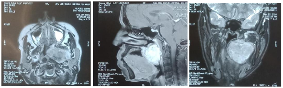
Figure 1: Magnetic resonance imaging showing a large left cervical mass and its relationship with adjacent anatomical structures in axial, sagittal, and coronal planes.
Surgical management:
The patient underwent surgical excision through a left transcervical approach. Following exposure, the lesion appeared as a giant multiloculated cystic mass occupying the left cervical compartment and extending deeply toward the thoracic inlet. Because of its considerable size and close relationship with the major cervical vessels, controlled aspiration of abundant serous fluid was performed to decrease the tumor volume and facilitate meticulous dissection. Following decompression, complete excision of the lesion was achieved while preserving the carotid artery, internal jugular vein, vagus nerve, spinal accessory nerve, and surrounding cervical structures. Macroscopic examination of the surgical specimen demonstrated a multiloculated cystic mass with thick fibrous septa and focal solid areas within the cyst wall (Figure 2).
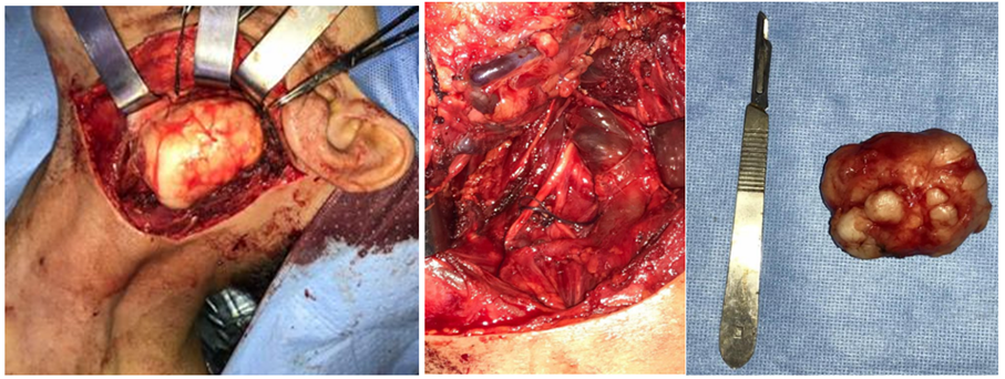
Figure 2: Intraoperative findings showing the giant multiloculated cervical mass and the surgical bed after complete excision.
Histopathological findings:
Microscopic examination revealed a malignant small round blue cell tumor composed of uniform round cells arranged in diffuse sheets. The tumor cells exhibited hyperchromatic nuclei, finely dispersed chromatin, inconspicuous nucleoli, and scant eosinophilic cytoplasm. Immunohistochemical analysis demonstrated diffuse membranous positivity for CD99 together with (NKX2.2 / FLI-1 selon tes résultats), supporting the diagnosis of primary extraskeletal Ewing sarcoma. Molecular analysis (FISH or RT-PCR for EWSR1 rearrangement, if performed) further confirmed the diagnosis.
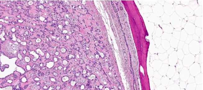
Figure 3: Histopathological features of extraskeletal Ewing sarcoma showing proliferation of uniform small round cells (H&E staining).
Postoperative management and follow-up:
Following histopathological confirmation, the case was discussed at a multidisciplinary tumor board including head and neck surgeons, pathologists, radiologists, medical oncologists, and radiation oncologists. The patient was referred for multimodal oncological treatment consisting of (à compléter selon le protocole : VDC/IE chemotherapy ± radiotherapy). At the latest follow-up (à compléter : X months), the patient remained (disease-free / under treatment / without evidence of local recurrence or distant metastasis).
Discussion
Extraskeletal Ewing sarcoma (EES) is a rare, highly aggressive malignant mesenchymal neoplasm belonging to the Ewing sarcoma family of tumors (ESFT). It accounts for approximately 15–20% of all Ewing sarcomas and shares the same histopathological, immunophenotypic, and molecular characteristics as conventional osseous Ewing sarcoma [1–3]. Although EES may occur at virtually any anatomical site, it predominantly arises in the deep soft tissues of the trunk, paravertebral region, pelvis, and lower extremities. Primary involvement of the head and neck is distinctly uncommon, representing only a small proportion of reported cases, while cervical localization remains exceptionally rare [2,4,5].
The rarity of cervical EES represents a major diagnostic challenge because its clinical presentation is usually nonspecific. Most patients present with a progressively enlarging painless cervical mass, whereas pain, neurological deficits, dysphagia, dysphonia, or respiratory symptoms generally occur only after compression of adjacent neurovascular or aerodigestive structures [4–7]. Our patient presented with an isolated rapidly enlarging cervical swelling without constitutional symptoms or neurological impairment despite the considerable tumor size. This relatively silent clinical course probably reflects the ability of the lesion to expand progressively along the cervical fascial planes before producing compressive manifestations.
Radiological assessment plays a central role in determining tumor extent and surgical planning but lacks sufficient specificity to establish a definitive diagnosis. In most published reports, cervical EES appears as a poorly or well-defined heterogeneous solid soft-tissue mass showing variable contrast enhancement with areas of necrosis or hemorrhage [2,5,8]. MRI usually demonstrates an isointense lesion on T1-weighted images and heterogeneous hyperintensity on T2-weighted sequences, with marked diffusion restriction owing to its high cellularity [8]. However, these findings overlap considerably with those observed in other malignant soft-tissue tumors, making imaging alone insufficient for diagnosis.
The radiological appearance observed in our patient was particularly unusual. CT demonstrated a giant multiloculated predominantly cystic lesion containing thick internal septa and only limited mural soft-tissue components. Moreover, surgical exploration confirmed multiple interconnected cystic cavities filled with abundant serous fluid, necessitating controlled aspiration before complete excision. Such a presentation has only exceptionally been reported in the literature and substantially broadened the differential diagnosis. Indeed, both the imaging and intraoperative findings strongly suggested a benign congenital cystic lesion rather than a malignant mesenchymal tumor. This observation emphasizes that EES should not be excluded solely because of a predominantly cystic appearance, particularly in young adults presenting with rapidly enlarging cervical masses [4–8].
The differential diagnosis of a multiloculated cystic lateral neck mass is broad and includes second branchial cleft cysts, cystic lymphatic malformations, infected congenital cysts, cystic schwannomas, abscesses, necrotic metastatic lymphadenopathy, and, less frequently, cystic salivary gland neoplasms [5,8]. Clinical history, patient age, imaging characteristics, and growth kinetics may orient the diagnosis; nevertheless, considerable overlap exists between these entities. In the present case, the rapid increase in tumor size over only two months together with the presence of mural soft-tissue nodules represented subtle imaging findings suggestive of an underlying malignant process despite the predominantly cystic morphology.
Histopathological examination remains the cornerstone of diagnosis. Microscopically, EES consists of sheets of uniform small round blue cells with hyperchromatic nuclei, finely dispersed chromatin, inconspicuous nucleoli, and scant cytoplasm rich in glycogen [1,9]. Because these features overlap with several other malignant small round cell tumors, including rhabdomyosarcoma, lymphoma, poorly differentiated synovial sarcoma, mesenchymal chondrosarcoma, desmoplastic small round cell tumor, and neuroblastoma, immunohistochemical and molecular analyses are indispensable [9–11]. Diffuse membranous expression of CD99 remains the most sensitive immunohistochemical marker, while NKX2.2 and FLI-1 significantly improve diagnostic specificity. Whenever feasible, demonstration of an EWSR1 rearrangement by FISH or RT-PCR remains the diagnostic gold standard [10,11]. In our patient, the diagnosis was established only after complete surgical excision, highlighting the limitations of clinical and radiological evaluation in atypical presentations.
Current treatment recommendations advocate multimodal therapy combining systemic chemotherapy with complete surgical excision whenever feasible, followed by radiotherapy according to surgical margins, histological response, and tumor stage [12–15]. Neoadjuvant chemotherapy has become the standard approach in many specialized sarcoma centers because it facilitates tumor downsizing, eradicates occult micrometastatic disease, and provides valuable prognostic information through assessment of histological response [12]. However, surgery remains the cornerstone of local control in resectable lesions. In the head and neck region, complete resection may be technically demanding because of the intimate relationship between the tumor and major vessels, cranial nerves, and the upper aerodigestive tract. In the present case, controlled aspiration of the cystic content substantially reduced tumor volume and allowed meticulous dissection while preserving all major cervical neurovascular structures, thereby facilitating complete excision without intraoperative complications.
Several prognostic factors have been consistently associated with survival in patients with Ewing sarcoma, including metastatic disease at diagnosis, tumor size, axial location, histological response to chemotherapy, surgical margin status, and local recurrence [12–15]. Patients with localized disease treated using contemporary multimodal protocols currently achieve 5-year overall survival rates approaching 70–80%, whereas the presence of metastatic disease at diagnosis remains associated with a markedly poorer prognosis [13–15].
Finally, our case expands the clinicoradiological spectrum of cervical extraskeletal Ewing sarcoma. To our knowledge, reports describing a giant predominantly multiloculated cystic cervical EES containing abundant serous fluid and closely mimicking a congenital branchial cleft cyst remain exceptionally scarce. This unusual presentation underscores the importance of maintaining a high index of suspicion when evaluating rapidly enlarging cystic neck masses in young adults. Early histopathological diagnosis, multidisciplinary management, and prompte initiation of oncological treatment remain essential for improving clinical outcomes.
Conclusion
Primary cervical extraskeletal Ewing sarcoma is an exceptionally rare and highly aggressive malignancy that may present with atypical clinical and radiological features, posing a significant diagnostic challenge. Our case illustrates an unusual presentation as a giant multiloculated predominantly cystic neck mass closely mimicking a congenital branchial cleft cyst both radiologically and intraoperatively. This observation highlights that malignancy should always be considered in the differential diagnosis of rapidly enlarging complex cystic cervical lesions, particularly in young adults. Histopathological examination complemented by immunohistochemistry and molecular analysis remains essential for establishing the diagnosis, while optimal outcomes rely on prompt multidisciplinary management integrating surgery and systemic oncological treatment. Reporting additional cases is necessary to better define the clinicopathological spectrum, optimize diagnostic strategies, and improve therapeutic management of this exceptionally rare entity.
Declarations
Ethics approval and consent to participate: Ethical approval was not required for this case report in accordance with our institutional policy.
Consent for publication: Written informed consent was obtained from the patient for publication of this case report and the accompanying images. A copy of the written consent is available for review by the Editor-in-Chief of this journal upon reasonable request.
Availability of data and materials: All data generated or analyzed during this study are included in this published article. Additional data are available from the corresponding author upon reasonable request.
Competing interests: The authors declare that they have no competing interests.
Funding: The authors received no specific funding for this work.
Authors’ contributions
F.E.M. contributed to patient management, study conception, data collection, literature review, manuscript drafting, and manuscript submission.
M.L. (Meriem Lahjaouj), M.L. (Meriem Loudghiri), W.B., Y.O., S.R., R.A., M.R., and M.M. contributed to patient management, critical revision of the manuscript, and supervision of the study.
All authors read and approved the final manuscript.
Acknowledgements: The authors would like to thank the Department of Otorhinolaryngology–Head and Neck Surgery, CHU Ibn Rochd, Casablanca, Morocco, for its support in the diagnosis and management of this patient.
References
- Grünewald TGP, Cidre-Aranaz F, Surdez D, Tomazou EM, de Álava E, Kovar H, et al. Ewing sarcoma. Nat Rev Dis Primers, 2018; 4: 5.
- Delattre O, Zucman J, Melot T, Garau XS, Zucker JM, Lenoir GM, et al. The Ewing family of tumors—a subgroup of small-round-cell tumors defined by specific chimeric transcripts. N Engl J Med, 1994; 331(5): 294-299.
- Applebaum MA, Worch J, Matthay KK, Goldsby R, Neuhaus J, West DC, et al. Clinical features and outcomes in patients with extraskeletal Ewing sarcoma. Cancer, 2011; 117(13): 3027-3032.
- Almohaisen GA, Alhuwairini SF, Aljrayed MA, Alenezi M, Alsaab F. Extraskeletal Ewing's sarcoma of the head and neck region in pediatric patients: A case report and literature review. Int J Surg Case Rep, 2023; 106: 108142.
- Ali S, Mackenzie K, Reid R, O'Neill G. Cervical extraskeletal Ewing's sarcoma: Case report demonstrating radiological features and management. J Laryngol Otol, 2008; 122(9): 998-1001.
- Jakovljević S, Arsović N, Dudvarski Z, et al. Primary cervical extraosseous Ewing sarcoma originated from the sternocleidomastoid muscle: A case report and review of the literature. Case Rep Otolaryngol, 2024; 2024: 8867131.
- Wang S, Zhu W, Zhang H, Yang X. Extraosseous Ewing sarcoma of the cervical esophagus: A case report and literature review. Ear Nose Throat J, 2022; 101(10): NP214-NP219.
- Murphey MD, Senchak LT, Mambalam PK, Logie CI, Klassen-Fischer MK, Kransdorf MJ. From the radiologic pathology archives: Ewing sarcoma family of tumors: Radiologic-pathologic correlation. Radiographics, 2013; 33(3): 803-831.
- Machado I, Navarro S, Picci P, Llombart-Bosch A. Ewing sarcoma and the new emerging Ewing-like sarcomas: (CIC and BCOR-rearranged sarcomas). Semin Diagn Pathol, 2016; 33(1): 10-19.
- Yoshida A, Sekine S, Tsuta K, Fukayama M, Furuta K, Tsuda H. NKX2.2 is a useful immunohistochemical marker for Ewing sarcoma. Am J Surg Pathol, 2012; 36(7): 993-999.
- WHO Classification of Tumours Editorial Board. Soft Tissue and Bone Tumours. 5th ed. Lyon: International Agency for Research on Cancer, 2020.
- Gaspar N, Hawkins DS, Dirksen U, Lewis IJ, Ferrari S, Le Deley MC, et al. Ewing sarcoma: Current management and future approaches through collaboration. J Clin Oncol, 2015; 33(27): 3036-3046.
- Grier HE, Krailo MD, Tarbell NJ, Link MP, Fryer CJ, Pritchard DJ, et al. Addition of ifosfamide and etoposide to standard chemotherapy for Ewing's sarcoma and primitive neuroectodermal tumor of bone. N Engl J Med, 2003; 348(8): 694-701.
- Leavey PJ, Mascarenhas L, Marina N, Chen Z, Krailo M, Miser J, et al. Prognostic factors for patients with Ewing sarcoma (Ewing's Sarcoma Family of Tumors) treated on pediatric oncology group protocols. J Clin Oncol, 2008; 26(27): 4385-4393.
- Balamuth NJ, Womer RB. Ewing's sarcoma. Lancet Oncol, 2010; 11(2): 184-192.