
Email id: info@ijclinmedcasereports.com
Inflammatory Fibroid Polyp of the Ileum Presenting as Adult Intussusception and Small Bowel Obstruction in an Elderly Patient: A Case Report and Literature Review
Mohamed Fadil*, Mehdi Salmane, Asmae Guennouni, Rachida Saouab, Jamal El Fenni and Zakaria Toufga
Radiology department, Mohamed V military hospital, Rabat, Morocco
Received Date: 25/08/2026; Published Date: 28/10/2026
*Corresponding author: Mohamed Fadil, Radiology department, Mohamed V military hospital, Rabat, Morocco
Abstract
Inflammatory fibroid polyps (IFPs), or Vanek’s tumors, are rare, benign gastrointestinal mesenchymal neoplasms. While predominantly found in the stomach, small bowel localization causing acute mechanical occlusion via intussusception is exceptionally rare. We present the case of a 76-year-old female with an acute small bowel obstruction due to ileo-ileal intussusception induced by an ileal IFP, confirmed through imaging, emergency exploratory laparotomy, and histopathological evaluation with positive CD34 immunohistochemistry.
Keywords: Inflammatory fibroid polyp; Vanek’s tumor; Small bowel obstruction; Ileal intussusception; Case report
Introduction
Inflammatory Fibroid Polyps (IFPs), originally characterized by Vanek in 1949 under the nomenclature of "gastric submucosal granulomas with eosinophilic infiltration," are uncommon, non-neoplastic benign lesions derived from the mesenchymal elements of the gastrointestinal tract submucosa [1]. Although the stomach represents the most prevalent anatomical site, specifically the gastric antrum—IFPs can theoretically develop anywhere along the length of the alimentary canal, with the small intestine, and more specifically the ileum, representing a distinctly uncommon location [1,2].
Adult intussusception accounts for a mere fraction of mechanical bowel obstructions, representing roughly 5% of all cases, and is fundamentally characterized by an underlying organic pathological lead point, such as a neoplasm, Meckel’s diverticulum, or stricture. While small bowel IFPs may remain quiescent and clinically silent for extended periods, progressive radial expansion frequently transforms them into mechanical lead points that trigger localized intussusception, ultimately culminating in subocclusive or acute complete mechanical bowel obstruction [3,4].
This article details a rare clinical presentation of ileal occlusion secondary to an IFP-induced intussusception in a 76-year-old patient, correlating pre-operative imaging findings and intraoperative parameters with definitive histopathological confirmation, and contextualizes the case within the contemporary medical literature.
Case Presentation
A 76-year-old female presented to our department with a one-week history of persistent, vomiting and progressive abdominal pain, accompanied by obstipation (absence of stool emission) for 24 hours prior to admission. Upon clinical evaluation, the patient remained hemodynamically, respiratory, and neurologically stable.
An urgent contrast-enhanced abdominal and pelvic CT scan was performed, which demonstrated stomach and proximal small bowel distension (predominantly jejunal and upper ileal) with fluid-gas levels upstream of a distinct transition zone. Cross-sectional imaging identified an oval-shaped, well-circumscribed, hypodense polypoid mass measuring mm, obstructing the lumen of the proximal ileum, showing delayed enhancement post-contrast and a visible pedicle attaching it to the anterior bowel wall. Distal ileal loops and the colon were collapsed. A small peritoneal effusion was noted in the left iliac fossa extending into the pelvis, indicating early bowel distress without evidence of transmural necrosis, pneumatosis, or pneumoperitoneum (Figure 1).
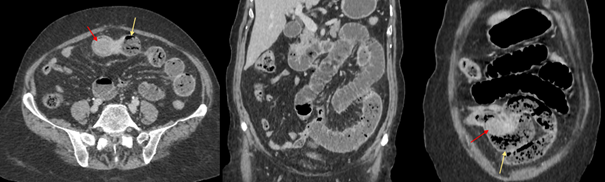
Figures 1: Contrast-enhanced abdominal CT scan (Axial and Coronal views), demonstrates marked proximal intestinal distension (predominantly jejunal) with fluid-gas levels and a distinct "small bowel feces sign" (Yellow arrow) immediately proximal to a well-defined, obstructive intraluminal polypoid mass serving as the lead point (Red arrow).
The patient was taken to the operating room for an emergency exploratory laparotomy. Intraoperative exploration confirmed high-grade mechanical small bowel obstruction secondary to localized invagination (intussusception) of the proximal ileum. Manual reduction exposed a firm, pedunculated intraluminal polyp acting as the mechanical lead point (Figure 2). A segmental small bowel resection was performed, followed by a primary intestinal restoration.
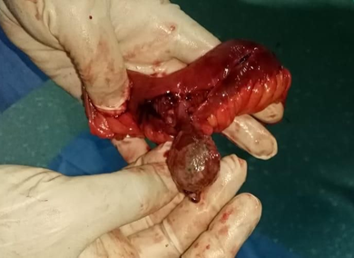
Figure 2: Intraoperative surgical photograph, shows the exteriorized small bowel segment following enterectomy, demonstrating the firm, pedunculated intraluminal polypoid formation acting as the mechanical lead point for the intussusception.
Gross pathological evaluation of the resected small bowel specimen (16 x 3 cm) revealed a well-formed pedunculated polyp measuring 4 x 3 x 2.5 cm, located from the nearest surgical margin. Histopathological examination confirmed a submucosal stromal cell proliferation with capillary hypervascularity, a dense eosinophilic infiltrate, and strong positive anti-CD34 immunoreactivity. These findings established the definitive diagnosis of a benign ileal inflammatory fibroid polyp (Vanek’s tumor) with no evidence of malignancy.
The patient experienced an uncomplicated postoperative course and was successfully discharged following complete recovery of gastrointestinal function.
Discussion
Inflammatory fibroid polyps remain a subject of significant diagnostic and pathogenetic interest within gastrointestinal pathology. While historically classified as reactive or inflammatory pseudotumors driven by local physical, chemical, or infectious stimuli, modern molecular pathology has fundamentally shifted this perspective [1]. The discovery of activating mutations within the gene encoding the platelet-derived growth factor receptor alpha (PDGFRA) in a substantial proportion of IFPs has firmly established their status as true neoplastic mesenchymal proliferations rather than purely reactive processes [1,5].
Demographically, IFPs demonstrate a broad age distribution but exhibit a notable peak incidence between the fifth and seventh decades of life [1]. The presentation of an IFP in an elderly patient carries heightened clinical risk. Geriatric patients possess reduced physiological reserve and an elevated baseline susceptibility to rapid clinical deterioration, including bowel ischemia, transmural necrosis, and secondary peritonitis, if an acute mechanical occlusion is not promptly diagnosed and surgically managed [1,4].
Small bowel IFPs possess a distinct propensity for precipitating acute abdominal crises. As documented across multiple clinical series and case reports, polyps arising within the jejunum or ileum rarely present with insidious dyspeptic symptoms or chronic anemia; instead, due to the narrower caliber of the small bowel compared to the stomach, they rapidly manifest with subocclusive syndromes or acute mechanical obstruction driven by intussusception [2-4,6]. Unlike gastric IFPs, which are frequently amenable to diagnostic and therapeutic endoscopic mucosal resection or snare polypectomy during routine esophagogastroduodenoscopy, small bowel lesions remain occult until complications arise [1]. Consequently, cross-sectional imaging via contrast-enhanced CT is indispensable, serving as the gold standard modality for identifying the pathognomonic target sign, localizing the mechanical lead point, and staging associated bowel wall distress [1,4].
Histopathologically, the diagnosis relies on recognizing the classic triad of a submucosal stromal proliferation of spindle cells, a prominent network of blood vessels with concentric perivascular cuffing, and a heavy infiltration of eosinophils [4,6]. Although the vast majority of IFPs remain strictly confined within the submucosal layer, rare cases exhibiting deep tissue invasion extending into the muscularis propria or subserosal layers have been reported in contemporary literature, challenging the traditional dogma of purely localized disease and further supporting their neoplastic nature [5]. Furthermore, atypical immunohistochemical profiles—such as occasional focal loss or negativity for CD34, which is typically a reliable diagnostic adjunct—can complicate pathological interpretation, necessitating meticulous correlation with morphological features [5].
Ultimately, because small bowel IFPs can clinically and radiologically mimic malignant mesenchymal neoplasms such as Gastrointestinal Stromal Tumors (GISTs) or adenocarcinomas, definitive surgical resection remains both the primary therapeutic intervention for acute obstruction and the definitive diagnostic modality.
Conclusion
Small bowel inflammatory fibroid polyps are rare, benign mesenchymal neoplasms that can precipitate dramatic acute surgical emergencies, including intussusception and high-grade mechanical occlusion, particularly in elderly populations. Preoperative computed tomography is crucial for identifying the characteristic intussusception and mechanical lead point. Prompt surgical resection provides a definitive cure while eliminating diagnostic uncertainty regarding potential malignant mimics.
References
- Abboud B. Vanek’s tumor of the small bowel in adults. World Journal of Gastroenterology, 2015; 21(16): 4802. https://doi.org/10.3748/wjg.v21.i16.4802
- Sánchez, et al. Inflammatory Fibroid Polyp (Vanek’s Tumor): A Retrospective Multicentric Analysis of 67 Cases. Multicentric Medical Review, 2025.
- Ahtil R, et al. Etiologie rare d'un syndrome subocclusif: polype fibrinoïde inflammatoire de l'iléon, à propos d'un cas clinique. Pan African Medical Journal, 2017 ; 26 : 146. https://doi.org/10.11604/pamj.2017.26.146.10775
- Dagistanli, et al. A Rare Cause of Colonic Obstruction: Inflammatory Fibroid Polyp. Case Reports in Surgery, 2022.
- Tang, et al. Rare invasive inflammatory fibroid polyp presenting as small bowel intussusception: Two case reports and review of the literature, 2025.
- Toydemir T. Inflammatory fibroid polyp of the ileum presenting with small bowel obstruction in an adult patient: a case report. Journal of Medical Case Reports, 2010 ; 4 : 291. https://doi.org/10.1186/1752-1947-4-291.