
Email id: info@ijclinmedcasereports.com
Isolated myodesopsia revealing a solitary Choroidal Metastasis in a presumed cured Breast Cancer Patient: A Multimodal Imaging Diagnosis
Boulanouar Abdelilah1,*, Achour Inasse1, Aziz Anas1, Boutimzine Nourredine2 and Cherkaoui Lalla Ouafae2
1Department of Ophthalmology, Ibn Sina University Hospital, Rabat, Morocco
2Professor, Department of Ophthalmology, Ibn Sina University Hospital, Rabat, Morocco
Received Date: 12/08/2026; Published Date: 23/10/2026
*Corresponding author: Dr. Boulanouar Abdelilah, MD, Department of Ophthalmology, Speciality Hospital, Ibn Sina University Hospital, Rabat, Morocco
Abstract
Choroidal metastasis is the most frequent intraocular malignancy in adults, predominantly originating from primary breast carcinoma in women. Late ocular metastasis presenting years after complete oncological remission remains a diagnostic challenge. We report the case of a 54-year-old female with a history of invasive ductal carcinoma of the breast, declared in complete remission for three years, who presented with isolated myodesopsia (floaters) in her left eye. Standard ophthalmic evaluation revealed a creamy yellow-white subretinal mass superonasal to the optic disc. Diagnosis was established non-invasively using multimodal imaging: spectral-domain optical coherence tomography (SD-OCT) demonstrated a localized choroidal elevation with subretinal fluid; Fluorescein Angiography (FA) showed early hypofluorescence with late pinhole leakage; and B-scan ultrasonography confirmed a solid, dome-shaped mass (2.9*9.4 mm thickness) with medium-to-high internal reflectivity without choroidal excavation. The patient was referred for urgent systemic restaging but was lost to follow-up. Non-specific symptoms like floaters can herald late metastatic relapse, emphasizing the pivotal role of multimodal imaging.
Keywords: Choroidal metastasis; Breast carcinoma; Myodesopsia; Optic disc; Multimodal imaging
Introduction
The vascular network of the posterior uvea makes the choroid the primary intraocular site for hematogenous metastatic seeding [1]. Breast carcinoma accounts for 40% to 50% of choroidal metastases in women [2]. A major diagnostic hurdle in ocular oncology is the prolonged latency period between primary tumor treatment and choroidal metastasis, which can manifest years after complete oncological remission [3]. Although central visual loss is common, initial subtle presentations like isolated myodesopsia (floaters) are frequently overlooked [4]. Multimodal imaging—comprising spectral-domain optical coherence tomography (SD-OCT), fluorescein angiography (FA), and B-scan ultrasonography—provides pathognomonic diagnostic criteria that allow definitive non-invasive confirmation without intraocular biopsy [5]. We report a case of late choroidal metastasis superonasal to the optic disc in the left eye presenting as isolated myodesopsia, confirmed via multimodal imaging.
Case Report
A 54-year-old female presented with a three-week history of persistent myodesopsia (floaters) in her left eye, without photopsia, visual field loss, or pain. Eight years prior, she was diagnosed with invasive ductal carcinoma of the left breast (T2N1M0), treated with mastectomy, chemotherapy, radiotherapy, and five years of endocrine therapy, remaining in complete remission for three years.
Best-corrected visual acuity was 20/20 in the right eye and 20/25 in the left eye. Slit-lamp anterior segment examination was normal bilaterally. Fundoscopy of the left eye revealed an elevated, creamy yellow-white subretinal mass located superonasal to the optic disc (papilla) with overlying RPE alterations and localized shallow serous detachment. The right eye was normal.
Multimodal imaging of the left eye demonstrated:
- SD-OCT: Dome-shaped choroidal elevation superonasal to the papilla with photoreceptor disruption, and subretinal fluid with hyperreflective lipofuscin debris.
- FA: Early-phase hypofluorescence followed by late-phase hyperfluorescent pinhole leakage and diffuse dye staining.
- B-scan Ultrasonography: A solid, dome-shaped mass (thickness 2.4 mm, basal diameter: 9.4 mm) with medium-to-high internal reflectivity and no choroidal excavation.
A clinical diagnosis of choroidal metastasis secondary to breast carcinoma was established. The patient was referred for systemic oncology restaging and multidisciplinary management; however, she was subsequently lost to follow-up despite multiple contact attempts.
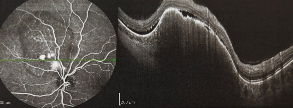
Figure 1: SD-OCT of the Left Eye. Cross-sectional scan through the lesion superonasal to the papilla showing dome-shaped elevation with "lumpy-bumpy" contour, photoreceptor disruption, and subretinal fluid.
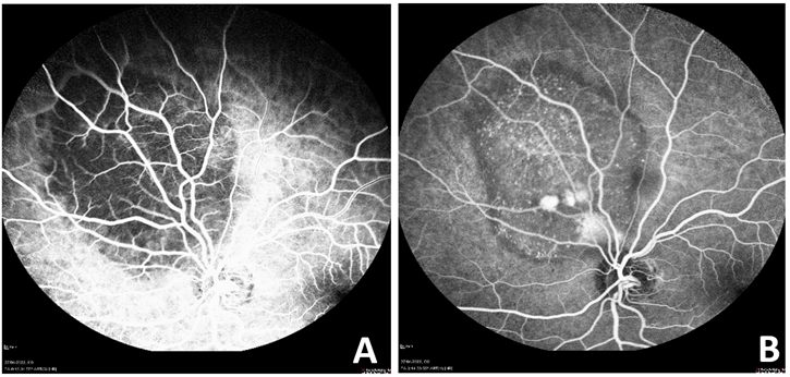
Figure 2: Fluorescein Angiography of the Left Eye. (A) Early phase showing patchy hypofluorescence superonasal to the disc. (B) Late phase revealing pinhole leakage and diffuse staining.
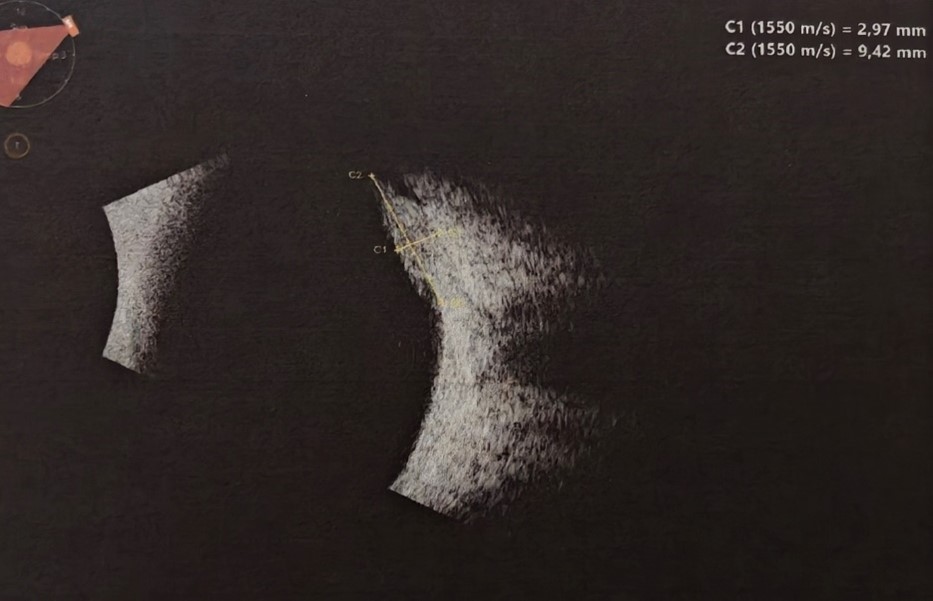
Figure 3: B-Scan Ultrasonography of the Left Eye. Echogram showing a solid mass superonasal to the papilla (thickness: 2.9* 9.4 mm) with medium-to-high internal reflectivity and no choroidal excavation.
Discussion
Choroidal metastasis presenting as isolated floaters superonasal to the papilla in a long-term survivor of breast cancer poses a significant diagnostic pitfall [1,4]. Extended latency reflects cellular tumor dormancy, where micro-metastatic clones persist in a quiescent state before re-activating [3]. Myodesopsia in this setting stems from vitreous micro-seeding, localized vitreopapillary traction, or optical distortion from subretinal fluid [5]. Preserved visual acuity (20/25) due to the extrafoveal location can cause missed diagnoses if dilated fundus evaluation is omitted [6].
Multimodal imaging is essential for differentiation from other intraocular tumors [6,7]:
- SD-OCT: Shows a "lumpy-bumpy" choroidal surface and subretinal fluid, contrasting with the smoother profiles of melanomas or hemangiomas [6].
- FA: Demonstrates early hypofluorescence and late pinhole leakage, unlike the rapid pre-arterial filling of hemangiomas or double circulation of melanomas [7].
- B-scan: Reveals medium-to-high internal reflectivity without choroidal excavation, distinguishing it from melanomas which display low reflectivity and acoustic hollowing [8].
Interdisciplinary management combining systemic therapy and local radiation is paramount [9,10]. Patient loss to follow-up in ocular oncology carries severe risks of irreversible visual loss, secondary glaucoma, and unchecked systemic progression [9].
Conclusion
New-onset myodesopsia can herald late choroidal metastasis superonasal to the optic disc in patients with a history of breast carcinoma. Non-invasive multimodal imaging enables rapid diagnostic confirmation. Robust patient-tracking systems are imperative to prevent loss to follow-up and ensure timely treatment.
Author Contributions :
Boulanouar, I.Achour, A. Aziz: Concept, design, acquisition of data, drafting, Guarantor of the work.
Pr N.Boutimzine, L.O. Cherkaoui: critical revision, and final approval of the version to be published.
Competing Interests: The author declares no competing interests.
Grant Information: The author received no specific funding for this work.
Acknowledgments: None.
References
- Shields CL, Welch RJ, Malik K, et al. Ocular metastasis: Analysis of 4220 tumors in 3104 patients. Ophthalmology, 2018; 125(11): 1815-1826.
- Demirci H, Shields CL, Chao AN, Shields JA. Uveal metastasis from breast cancer in 264 patients. Am J Ophthalmol, 2003; 136(2): 264-271.
- George B, Wirostko RD, Hanna E. Late manifestation of choroidal metastasis from breast carcinoma. Case Rep Ophthalmol Med, 2020; 2020: 8842109.
- Arepalli S, Kaliki S, Shields CL. Choroidal metastases: Origin, features, and systemic prognosis. Indian J Ophthalmol, 2015; 63(2): 122-127.
- Mathis T, Jardel P, Loria O, et al. New concepts in the diagnosis and management of choroidal metastases. Prog Retin Eye Res, 2019; 68: 144-176.
- Al-Dahmash SA, Shields CL, Kaliki S, et al. Enhanced depth imaging optical coherence tomography of choroidal metastasis. Retina, 2014; 34(8): 1588-1593.
- Amselem L, Cervera E, Diaz-Llopis M, et al. Multimodal imaging in choroidal metastases. Eur J Ophthalmol, 2011; 21(4): 431-438.
- Sobolewska B, Ziemssen F, Koch JM, et al. Choroidal metastases: Clinical features and multimodal imaging. BMC Ophthalmol, 2021; 21(1): 145.
- Konstantinidis L, Rospond-Kubiak I, Heimann H, et al. Management of choroidal metastases. Br J Ophthalmol, 2017; 101(3): 285-291.
- Shah SU, Mashayekhi A, Shields CL, et al. Photoreceptor changes in choroidal metastasis on SD-OCT. Ophthalmology, 2014; 121(1): 387-391.